 Back
BackIntroduction to the Cardiovascular System & Anatomy of the Heart
Study Guide - Smart Notes
 Tailored notes based on your materials, expanded with key definitions, examples, and context.
Introduction to the Cardiovascular System
Overview and Main Functions
The cardiovascular system (CV system) is essential for the transport of substances throughout the body. It consists of three main components: the heart, blood vessels, and blood. The primary function is to deliver oxygen and nutrients to cells, remove waste products, and transport hormones, immune cells, and clotting proteins to their target locations.
The Heart: Acts as the central pump.
Blood Vessels: Serve as conduits for blood flow.
Blood: The fluid medium, accounting for 7-8% of body weight.
Transport Functions: Oxygen and nutrients to cells; wastes to liver and kidneys; hormones and immune cells to targets.
Circuits of the Cardiovascular System
The CV system is divided into two main circuits: pulmonary and systemic. Each circuit serves a distinct function in gas exchange and nutrient delivery.
Pulmonary Circuit: Permits gas exchange in the lungs; supplied by the right heart.
Systemic Circuit: Transports blood to all tissues except the lungs; supplied by the left heart.
Blood Path: Blood leaves the right ventricle via pulmonary arteries, returns to the left atrium via pulmonary veins; systemic blood leaves the left ventricle via the aorta, returns to the right atrium via venae cavae.

Anatomy of the Heart
External and Internal Structure
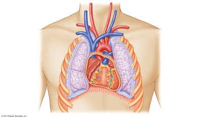
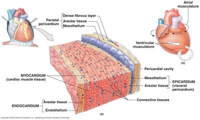
The heart is located in the thoracic cavity, separated from the abdominal cavity by the diaphragm. It is roughly the size of a fist and weighs 250–350 grams. The heart is surrounded by the pericardium, a membranous sac that lubricates and protects the heart.
Pericardium: Membranous sac; reduces friction.
Heart Wall Layers:
Epicardium: Outer layer; visceral pericardium.
Myocardium: Middle layer; muscular wall responsible for contraction.
Endocardium: Inner layer; epithelium providing protection for valves and chambers.
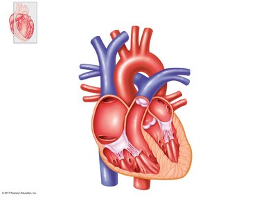
Heart Chambers and Valves
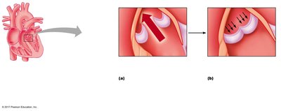
The heart contains four chambers: two atria and two ventricles. Valves ensure unidirectional blood flow, opening and closing in response to pressure gradients.
Atrioventricular (AV) Valves:
Right AV valve: Tricuspid valve
Left AV valve: Bicuspid (mitral) valve
Connected to myocardium by chordae tendineae and papillary muscles
Semilunar Valves:
Aortic valve (Aorta)
Pulmonary valve (Pulmonary Artery)
Valve Function: Prevent backward flow; open passively based on pressure gradient.
Wall Thickness of Ventricles
The left ventricle wall is thicker than the right ventricle wall due to the higher pressure required to pump blood throughout the systemic circuit, compared to the pulmonary circuit.
Left Ventricle: Pumps blood to systemic circulation; thicker wall.
Right Ventricle: Pumps blood to pulmonary circulation; thinner wall.
Cardiac Muscle Cells (Cardiocytes)
Types of Cardiocytes
Cardiac muscle cells are specialized for the heart's function. There are two main types: contractile and autorhythmic cells.
Contractile Cells:
Account for 99% of cardiocytes
Responsible for the pumping action
Small, bifurcate, single nucleus, aerobic, high in myoglobin and mitochondria
Extensive blood supply; involuntary contraction via sliding filament theory
Autorhythmic Cells:
Generate and spread action potentials spontaneously
Pacemaker cells: Initiate APs and establish heart rate
Conduction fibers: Transmit APs
Key difference from skeletal muscle: autorhythmicity
Characteristics of Cardiac Muscle
Cardiac muscle shares similarities with skeletal muscle but also has unique features.
Similarities: Striated, contain sarcomeres
Differences: Short, wide T tubules; less SR; single nucleus; intercalated discs
Intercalated Discs:
Gap Junctions: Electrically connect adjacent cells; functional syncytium
Desmosomes: Provide structural support; chemical communication
Summary Table: Heart Wall Layers
Layer | Structure | Function |
|---|---|---|
Epicardium | Outer layer; visceral pericardium | Protects heart |
Myocardium | Middle layer; cardiac muscle tissue | Contraction; pumping action |
Endocardium | Inner layer; epithelium | Protects valves and chambers |
Key Equations
Pressure Gradient and Blood Flow
Blood flow is driven by pressure differences:
Blood flows from high pressure to low pressure.
Equation for blood flow:
Q: Blood flow
ΔP: Pressure difference
R: Resistance
Recap and Learning Outcomes
After studying this material, you should be able to:
Identify the major components of the cardiovascular system and their functions.
Describe the major structures of the heart and the path of blood flow.
Differentiate between contractile and autorhythmic cardiocytes.
