 Back
BackIntroduction to the Nervous System and Nervous Tissue
Study Guide - Smart Notes
 Tailored notes based on your materials, expanded with key definitions, examples, and context.
Nervous System Overview
Central and Peripheral Nervous Systems
The nervous system is divided into the Central Nervous System (CNS) and the Peripheral Nervous System (PNS). The CNS consists of the brain and spinal cord, which are responsible for integrating and processing information. The PNS includes cranial and spinal nerves that connect the CNS to the rest of the body, facilitating communication between the CNS and peripheral organs.
Brain: Contains approximately 100 billion neurons, located in the skull, and controls all body functions.
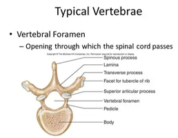
Spinal Cord: Contains about 100 million neurons, passes through the vertebral foramen, and connects the brain to the body below the head and neck.
Cranial Nerves: 12 pairs, connect directly to the brain.
Spinal Nerves: 31 pairs, connect to the spinal cord.
Functional Divisions of the Nervous System
Sensory, Integrative, and Motor Functions
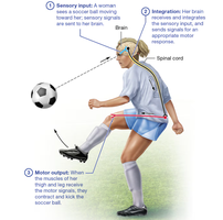
The nervous system performs three main functions:
Sensory Functions: Gathering information about internal and external environments.
Integrative Functions: Analyzing and interpreting sensory input.
Motor Functions: Initiating actions in response to integration, such as muscle contraction or gland secretion.
Nervous Tissue Structure
Neurons and Neuroglia
Nervous tissue is highly cellular, composed of neurons (excitable cells that transmit signals) and neuroglia (supporting cells). The extracellular matrix is minimal, and the tissue is specialized for rapid communication.
Neurons: Consist of a cell body, dendrites (receive input), and a single axon (transmits output).
Neuroglia: Support, protect, and maintain the environment around neurons; fill gaps when neurons die.
Neuron Structure and Function
Cell Body (Soma): Contains the nucleus and organelles; site of metabolic activity.
Dendrites: Branching processes that receive signals from other neurons; increase receptive surface area.
Axon: Single process that carries signals away from the cell body; may branch into axon collaterals and telodendria.
Axon Hillock: Region where the axon originates from the cell body.
Axolemma: Plasma membrane of the axon.
Axonal Transport
Slow Axonal Transport: Moves cytoskeletal proteins at 1-3 mm/day.
Fast Axonal Transport: Uses motor proteins and ATP to move materials at up to 200 mm/day.
Direction: Anterograde (toward axon terminal), Retrograde (toward cell body).
Functional Regions of a Neuron
Receptive Region: Dendrites and cell body; receive signals.
Conducting Region: Axon; transmits signals.
Secretory Region: Axon terminals; release neurotransmitters.
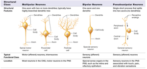
Structural Classes of Neurons
Neurons are classified based on the number and arrangement of their processes:
Structural Class | Features | Location |
|---|---|---|
Multipolar Neurons | One axon, two or more dendrites | Most neurons in CNS, motor neurons in PNS |
Bipolar Neurons | One axon, one dendrite | Special sensory organs (retina, olfactory epithelium) |
Pseudounipolar Neurons | Single process splits into two axons | Sensory neurons in PNS (touch, pain, vibration) |
Myelin Sheath and Nervous Tissue Types
Myelin Sheath
The myelin sheath is a lipid-rich insulating layer around axons, crucial for rapid signal conduction. Myelinated axons form white matter, while unmyelinated axons and cell bodies form gray matter.
Function: Prevents current leakage, increases conduction speed.
Unmyelinated Axons: Found in short axons of CNS and PNS; slower conduction.
Regeneration in the Nervous System
Steps of Axonal Regeneration
Distal axon and myelin sheath degenerate.
Growth processes form from the proximal axon.
Neurolemmocytes and basal lamina form a regeneration tube.
A single growth process enters the tube.
Axon reconnects with the target cell.
Electrophysiology of Neurons
Resting Membrane Potential and Ion Gradients
Neurons maintain a resting membrane potential due to differences in ion concentrations across the membrane, primarily sodium (Na+) and potassium (K+).
Electrochemical Gradient: Combination of electrical and chemical forces driving ion movement.
Leak Channels: Allow passive movement of ions, contributing to resting potential.
Local Potentials
Local (graded) potentials are small changes in membrane potential caused by a single stimulus. They can be:
Depolarization: Positive charges enter, making the membrane less negative.
Hyperpolarization: Positive charges exit or negative charges enter, making the membrane more negative.
Action Potentials
An action potential is a rapid, large change in membrane potential that propagates along the axon. It involves voltage-gated sodium and potassium channels cycling through resting, activated, and inactivated states.
Resting State: Inactivation gate open, activation gate closed.
Activated State: Both gates open.
Inactivated State: Inactivation gate closed, activation gate open.
Synaptic Transmission
Types of Synapses
Electrical Synapses: Direct current flow via gap junctions; bidirectional and nearly instantaneous.
Chemical Synapses: Use neurotransmitters released from synaptic vesicles into the synaptic cleft; more common and efficient.
Events at a Chemical Synapse
Action potential triggers opening of calcium channels in the presynaptic terminal.
Calcium influx causes vesicles to release neurotransmitters into the synaptic cleft.
Neurotransmitters bind to receptors on the postsynaptic neuron.
Ion channels open, generating a local potential and possibly an action potential.
Termination of Synaptic Transmission
Diffusion and absorption of neurotransmitter.
Degradation by enzymes in the synaptic cleft.
Reuptake into the presynaptic neuron.
Neurotransmitters and Their Functions
Major Neurotransmitter Classes
Acetylcholine: Involved in muscle contraction and autonomic functions.
Biogenic Amines (Monoamines): Include norepinephrine, epinephrine, dopamine, serotonin, and histamine; regulate mood, cognition, and autonomic functions.
Amino Acid Neurotransmitters: Glutamate (main excitatory NT in the brain), GABA (main inhibitory NT in the brain), Glycine (main inhibitory NT in the spinal cord).
Neuropeptides: Substance P (pain), opioids (pain relief), neuropeptide Y (hunger).
Neuromodulation
Some neurotransmitters act as neuromodulators, binding to metabolic receptors and triggering cascades that modulate neuron activity over longer periods.
Clinical Applications
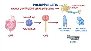
Poliomyelitis
Poliomyelitis is a viral infection that enters the CNS via retrograde axonal transport, causing severe nerve injury, paralysis, and potentially death. Other pathogens (herpes simplex, rabies, tetanus) use similar mechanisms.
Brain Fog and Myelin Loss
Microglia can reduce oligodendrocyte precursors and myelin, slowing impulse conduction and contributing to cognitive symptoms such as 'brain fog.'
Local Anesthetics
Local anesthetic drugs block sodium channels in the axolemma, preventing action potential generation and thus sensation of pain.
Psychiatric Disorders and Neurotransmitters
Schizophrenia: Excess dopamine; treated with dopamine receptor blockers.
Depressive Disorders: Deficiency in serotonin, norepinephrine, or dopamine; treated with SSRIs and related drugs.
Anxiety Disorders: Abnormal norepinephrine, serotonin, and GABA; treated with drugs enhancing GABA or modulating norepinephrine.
Bipolar Disorder: Treated with drugs that decrease axonal excitability, often by blocking sodium channels.
