 Back
BackIntroduction to the Nervous System and Nervous Tissue: Structured Study Notes
Study Guide - Smart Notes
 Tailored notes based on your materials, expanded with key definitions, examples, and context.
Overview of the Nervous System
Functions and Homeostasis
The nervous system is responsible for controlling perception, voluntary movement, consciousness, personality, learning, and memory. It works closely with the endocrine system to regulate homeostasis, including respiratory rate, blood pressure, body temperature, sleep/wake cycles, and blood pH.
Voluntary movement: Directs actions such as walking or speaking.
Homeostasis: Maintains internal balance through rapid signaling.
Integration: Processes sensory input and determines appropriate responses.
Anatomical Divisions of the Nervous System
Central and Peripheral Nervous Systems
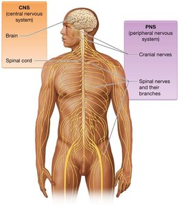
The nervous system is divided into two main anatomical divisions:
Central Nervous System (CNS): Consists of the brain and spinal cord. The brain contains billions of neurons and is protected by the skull. The spinal cord extends from the foramen magnum to the lumbar vertebrae, enabling communication between the brain and the body below the head and neck.
Peripheral Nervous System (PNS): Includes all nerves outside the CNS. Nerves are bundles of axons, blood vessels, and connective tissue. The PNS is further divided into cranial nerves (12 pairs) and spinal nerves (31 pairs).
Functional Divisions of the Nervous System
Sensory, Integrative, and Motor Functions
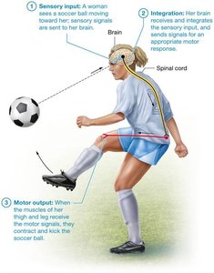
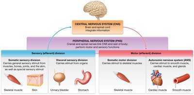
The nervous system is functionally categorized into three divisions:
Sensory (Afferent) Division: Gathers information from internal and external environments. Includes:
Somatic sensory division: Signals from muscles, bones, joints, skin, and special senses (vision, hearing, taste, smell, balance).
Visceral sensory division: Signals from internal organs (heart, lungs, stomach, kidneys, urinary bladder).
Integrative Functions: Analyze and interpret sensory information, determining appropriate responses. Most sensory input is subconsciously disregarded.
Motor (Efferent) Division: Carries out responses via motor neurons. Subdivided into:
Somatic motor division: Voluntary control of skeletal muscles.
Autonomic nervous system (ANS): Involuntary control of glands, smooth muscle, and cardiac muscle; critical for homeostasis.
Nervous Tissue
Neurons: Structure and Function
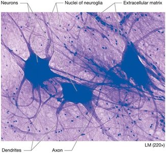
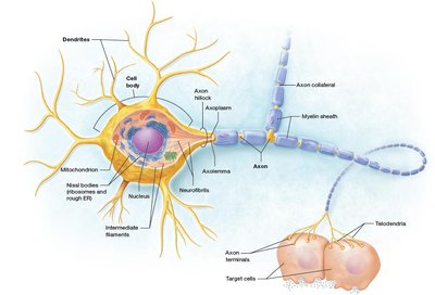
Neurons are excitable cells responsible for transmitting electrical signals (action potentials). Most neurons consist of three main parts:
Cell body (soma): Metabolically active, contains organelles for protein synthesis (ribosomes, rough ER/Nissl bodies), Golgi apparatus, nucleoli, mitochondria, and cytoskeleton (microtubules, neurofibrils).
Dendrites: Short, branched processes that receive input and transmit it toward the cell body.
Axon: Single, long process that conducts action potentials. Key regions include the axon hillock, axon collaterals, axon terminals (synaptic bulbs), axolemma (membrane), and axoplasm (cytoplasm).
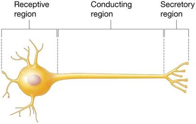
Functional Regions of Neurons
Receptive region: Dendrites and cell body.
Conducting region: Axon.
Secretory region: Axon terminal.
Neuron Classification
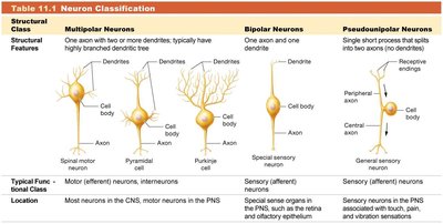
Neurons are classified by structural features and function:
Multipolar: One axon, multiple dendrites; most common.
Bipolar: One axon, one dendrite; found in eye and olfactory epithelium.
Pseudounipolar: Single fused axon with two branches; sensory neurons.
Sensory (afferent) neurons: Carry information toward CNS; usually pseudounipolar or bipolar.
Interneurons: Relay information within CNS; most abundant; multipolar.
Motor (efferent) neurons: Carry information from CNS to muscles/glands; mostly multipolar.
Structural Class | Features | Location | Function |
|---|---|---|---|
Multipolar | One axon, multiple dendrites | CNS, PNS | Motor, interneurons |
Bipolar | One axon, one dendrite | Eye, olfactory epithelium | Sensory |
Pseudounipolar | Single fused axon | PNS | Sensory |
Neuron Groupings
CNS: Nuclei (cell bodies), tracts (axons).
PNS: Ganglia (cell bodies), nerves (axons).
Neuroglia (Neuroglial Cells)
Types and Functions
Neuroglia provide structural support, protection, and environmental maintenance for neurons. They can divide and fill spaces left by dead neurons. There are six main types:
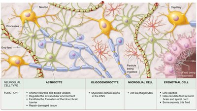
CNS:
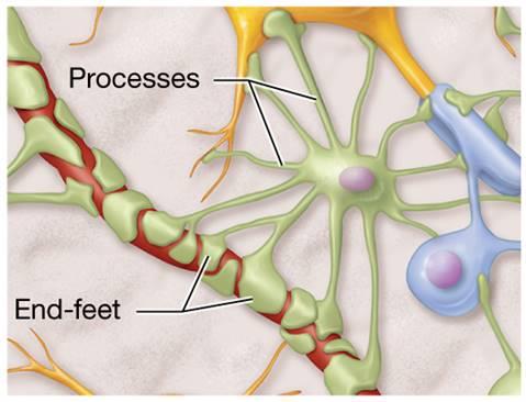
Astrocytes: Anchor neurons/blood vessels, regulate environment, form blood-brain barrier, repair tissue.
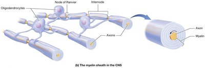
Oligodendrocytes: Myelinate axons.
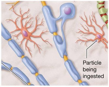
Microglia: Phagocytic, ingest debris/microorganisms.
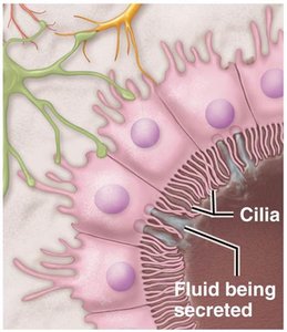
Ependymal cells: Ciliated, line CNS spaces, produce/circulate cerebrospinal fluid.
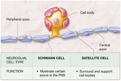
PNS:
Schwann cells: Myelinate axons.
Satellite cells: Support cell bodies.
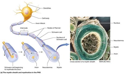
The Myelin Sheath
Structure and Function
The myelin sheath is formed by layers of plasma membrane from Schwann cells (PNS) or oligodendrocytes (CNS). Myelination insulates axons, increasing the speed of action potential conduction (15–20 times faster than unmyelinated axons).
Neurolemma: Present in PNS, absent in CNS.
Number of axons myelinated: Oligodendrocytes myelinate multiple axons; Schwann cells myelinate one axon.
Timing: Myelination begins earlier in PNS than CNS.
Internodes: Segments covered by myelin.
Node of Ranvier: Gaps between myelinated segments.

White and Gray Matter
White matter: Myelinated axons; appears white.
Gray matter: Cell bodies, unmyelinated dendrites/axons; appears gray.
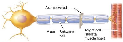
Regeneration of Nervous Tissue
Repair in CNS and PNS
Regeneration is nearly nonexistent in CNS, limited in PNS. Neural tissue can regenerate only if the cell body remains intact.
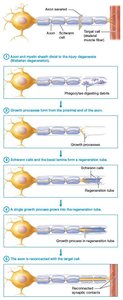
Steps:
Axon and myelin sheath degenerate distal to injury (Wallerian degeneration).
Growth processes form from proximal end.
Schwann cells and basal lamina form regeneration tube.
Growth process enters tube, directs new axon toward target.
New axon reconnects to target cell.
Electrophysiology of Neurons
Resting Membrane Potential
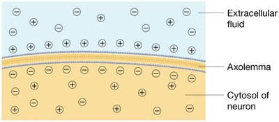
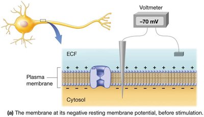
Neurons are excitable and respond to stimuli by generating electrical changes across their plasma membrane. The resting membrane potential (RMP) is typically −70 mV, due to the distribution of ions across the membrane.
RMP is maintained by leak channels (more K+ leaks out than Na+ leaks in) and the sodium-potassium pump.
Cell is polarized when voltage difference does not equal 0 mV.
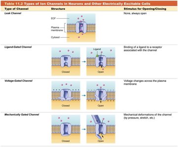
Ion Channels and Gradients
Leak channels: Always open.
Gated channels: Open in response to specific stimuli. Types include ligand-gated, voltage-gated, and mechanically-gated channels.
Sodium-potassium pump: Moves 3 Na+ out and 2 K+ in per ATP hydrolyzed, maintaining gradients.
Type of Channel | Structure | Stimulus |
|---|---|---|
Leak | Always open | None |
Ligand-gated | Opens with ligand binding | Chemical |
Voltage-gated | Opens with voltage change | Electrical |
Mechanically-gated | Opens with mechanical force | Pressure/stretch |
Changes in Membrane Potential
Depolarization: Positive charges enter, membrane potential becomes less negative.
Repolarization: Returns to resting potential.
Hyperpolarization: Membrane potential becomes more negative than at rest.
Local Potentials
Small, local changes in membrane potential; may cause depolarization or hyperpolarization.
Graded, reversible, and decremental (lose strength over distance).
Action Potentials
Phases and Mechanisms
Action potentials are rapid, uniform depolarizations and repolarizations generated in trigger zones (axon hillock, initial segment). They involve voltage-gated sodium and potassium channels.
Depolarization: Membrane potential rises toward zero, then becomes positive.
Repolarization: Returns to negative value.
Hyperpolarization: Temporarily more negative than resting potential.
All-or-None Principle
Action potentials occur only if threshold is reached (−55 mV).
Not dependent on stimulus strength, frequency, or length.
Irreversible and nondecremental (do not lose strength over distance).
Refractory Period
Absolute refractory period: No additional action potential can be generated.
Relative refractory period: Only strong stimulus can produce action potential.
Propagation of Action Potentials
Conduction and Speed
Action potentials are self-propagating, traveling in one direction from trigger zone to axon terminals.
Conduction speed depends on axon diameter and myelination.
Saltatory conduction: In myelinated axons, action potentials jump from node to node, increasing speed.
Continuous conduction: In unmyelinated axons, action potentials propagate along every section, slower.
Classification of Axons
Type A fibers: Fastest, largest diameter, myelinated; sensory/motor axons for skeletal muscle/joints.
Type B fibers: Intermediate speed/diameter, mostly myelinated; autonomic efferent fibers.
Type C fibers: Slowest, smallest diameter, unmyelinated; transmit pain, temperature, pressure.
Neuronal Synapses
Types and Structure
A synapse is the site where a neuron communicates with another cell. Synapses can be electrical (gap junctions) or chemical (neurotransmitter release).
Axodendritic: Axon to dendrite.
Axosomatic: Axon to cell body.
Axoaxonic: Axon to axon.
Electrical Synapses
Direct ion flow via gap junctions; rapid, synchronized activity (e.g., breathing, cardiac muscle).
Chemical Synapses
Most common; convert electrical signals to chemical signals via neurotransmitter release.
Contain synaptic vesicles, synaptic cleft, and neurotransmitter receptors.
Unidirectional; allow variable signal intensities.
Events at Chemical Synapse
Action potential triggers opening of voltage-gated calcium channels.
Calcium influx causes neurotransmitter release into synaptic cleft.
Neurotransmitter binds to postsynaptic receptors.
Ion channels open, leading to local potential and possibly action potential.
Termination of Synaptic Transmission
Neurotransmitter diffuses away, is degraded by enzymes, or reabsorbed (reuptake).
Major Neurotransmitters
Acetylcholine (ACh)
Widely used; binds cholinergic synapses in neuromuscular junction, brain, spinal cord, and ANS.
Largely excitatory; synthesized from choline and acetyl-CoA.
Degraded by acetylcholinesterase (AChE); by-products recycled.
Summary Table: Key Concepts
Concept | Key Points |
|---|---|
Nervous System Divisions | CNS (brain, spinal cord), PNS (nerves) |
Neuron Structure | Cell body, dendrites, axon |
Neuroglia Types | Astrocytes, oligodendrocytes, microglia, ependymal (CNS); Schwann, satellite (PNS) |
Myelin Sheath | Insulates axons, increases conduction speed |
Action Potential | Depolarization, repolarization, hyperpolarization; all-or-none |
Synapse | Electrical (gap junctions), chemical (neurotransmitters) |
Neurotransmitter | Acetylcholine (ACh), excitatory/inhibitory effects |
Key Equations
Sodium-Potassium Pump
The sodium-potassium pump maintains ion gradients:
Resting Membrane Potential
Typical neuron RMP:
Threshold for Action Potential
Threshold value:
Peak Depolarization
Peak of action potential:
Additional info:
These notes expand on brief points from slides and textbook images, providing definitions, examples, and context for college-level study.
Images included are directly relevant to the adjacent explanations, reinforcing anatomical and physiological concepts.
