 Back
BackIntroduction to the Nervous System and Nervous Tissue: Structured Study Notes
Study Guide - Smart Notes
 Tailored notes based on your materials, expanded with key definitions, examples, and context.
Overview of the Nervous System
General Functions
The nervous system is responsible for controlling perception, voluntary movement, consciousness, personality, learning, and memory. It also regulates homeostasis in conjunction with the endocrine system, affecting respiratory rate, blood pressure, body temperature, sleep/wake cycles, and blood pH.
Perception and Experience: The nervous system interprets sensory information and shapes our interaction with the environment.
Voluntary Movement: Directs skeletal muscle activity.
Homeostasis: Maintains internal balance through rapid signaling.
Anatomical Divisions
The nervous system is divided into the Central Nervous System (CNS) and Peripheral Nervous System (PNS).
CNS: Consists of the brain and spinal cord. The brain contains billions of neurons and is protected by the skull. The spinal cord extends from the foramen magnum to the lumbar vertebrae, enabling communication between the brain and the body.
PNS: Comprises all nerves outside the CNS, including cranial nerves (12 pairs) and spinal nerves (31 pairs). Nerves are bundles of axons with blood vessels and connective tissue.
Functional Divisions
The nervous system is functionally categorized into sensory, integrative, and motor divisions.
Sensory (Afferent) Division: Gathers information from internal and external environments via somatic (skeletal muscles, skin, special senses) and visceral (internal organs) subdivisions.
Integrative Functions: Analyze and interpret sensory input, determining appropriate responses. Most sensory information is subconsciously disregarded.
Motor (Efferent) Division: Executes responses via somatic (skeletal muscle, voluntary) and autonomic (smooth/cardiac muscle, glands, involuntary) subdivisions.
Nervous Tissue
Neurons: Structure and Function
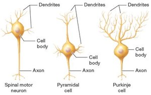
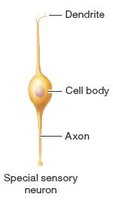
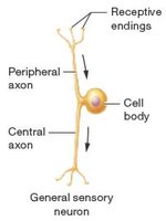
Neurons are excitable cells that transmit electrical signals (action potentials). Each neuron consists of three main parts:
Cell Body (Soma): Metabolically active region containing organelles for protein synthesis and energy production.
Dendrites: Short, branched processes that receive input and transmit it toward the cell body.
Axon: Single, long process that conducts action potentials away from the cell body. Includes axon hillock, axon collaterals, telodendria, axon terminals, and axolemma.
Axonal Transport
Substances move through the axon by slow (cytoskeletal proteins, 1–3 mm/day) or fast (vesicles, organelles, up to 400 mm/day) axonal transport, using motor proteins and ATP.
Functional Regions of Neurons
Receptive Region: Dendrites and cell body
Conducting Region: Axon
Secretory Region: Axon terminal
Neuron Classification
Structural: Multipolar (one axon, multiple dendrites), Bipolar (one axon, one dendrite), Pseudounipolar (one fused axon, two processes)
Functional: Sensory (afferent), Interneurons (association), Motor (efferent)
Neuron Groupings
CNS: Nuclei (cell bodies), Tracts (axons)
PNS: Ganglia (cell bodies), Nerves (axons)
Neuroglia
Neuroglial cells provide structural support, protection, and environmental maintenance for neurons.
CNS Types: Astrocytes (support, blood-brain barrier), Oligodendrocytes (myelination), Microglia (phagocytosis), Ependymal cells (cerebrospinal fluid)
PNS Types: Schwann cells (myelination), Satellite cells (support)
The Myelin Sheath
Structure and Function
The myelin sheath is formed by Schwann cells (PNS) or oligodendrocytes (CNS), wrapping layers of membrane around axons.
Insulation: Lipid-rich myelin prevents ion movement, increasing action potential speed.
Myelinated vs. Unmyelinated: Myelinated axons conduct signals 15–20 times faster.
Nodes of Ranvier: Gaps between myelinated segments where action potentials are regenerated.
White Matter: Myelinated axons
Gray Matter: Cell bodies, unmyelinated dendrites/axons
Electrophysiology of Neurons
Resting Membrane Potential
Neurons maintain a resting membrane potential (RMP) due to the distribution of ions across the plasma membrane.
Voltage: Electrical gradient from charge separation
Typical RMP: Slightly negative due to more potassium leaking out than sodium leaking in
Polarization: Cell is polarized when voltage difference is not zero
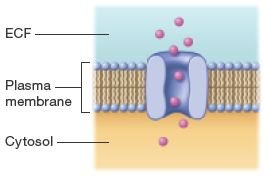
Ion Channels and Gradients
Ion movement across the membrane relies on specific channels:
Leak Channels: Always open, allow ions to flow down gradients
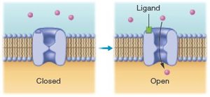
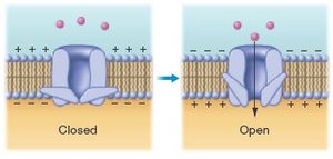
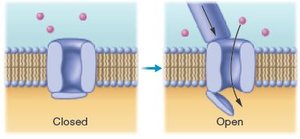
Gated Channels: Open in response to stimuli
Types: Ligand-gated (chemical), Voltage-gated (voltage), Mechanically-gated (mechanical)
Sodium-Potassium Pump
The sodium-potassium pump moves three sodium ions out and two potassium ions in per ATP hydrolyzed, maintaining gradients essential for RMP.
Changes in Membrane Potential
Depolarization: Positive charges enter, making membrane less negative
Repolarization: Return to RMP
Hyperpolarization: Membrane becomes more negative than RMP
Local Potentials
Local (graded) potentials are small, reversible changes in membrane potential, useful for short-distance signaling.
Depolarization: Moves membrane potential toward zero
Hyperpolarization: Moves membrane potential away from zero
Action Potentials
Action potentials are rapid, uniform depolarizations and repolarizations, generated in trigger zones.
Phases: Depolarization, repolarization, hyperpolarization
All-or-None Principle: Action potentials occur fully or not at all
Propagation: Action potentials travel from trigger zone to axon terminal, self-propagating in one direction
Conduction Speed: Influenced by axon diameter and myelination (saltatory vs. continuous conduction)
Classification of Axons
Type | Diameter | Myelination | Speed | Function |
|---|---|---|---|---|
Type A | Largest | Myelinated | Fastest (120 m/sec) | Sensory/motor for skeletal muscle and joints |
Type B | Intermediate | Mostly myelinated | Slower (15 m/sec) | Autonomic efferent, some sensory |
Type C | Smallest | Unmyelinated | Slowest (0.5–2 m/sec) | Pain, temperature, pressure |
Neuronal Synapses
Types of Synapses
Electrical Synapses: Direct ion flow via gap junctions; bidirectional and nearly instantaneous; found in programmed behaviors and muscle coordination.
Chemical Synapses: Use neurotransmitters; unidirectional; allow variable signal intensities; involve synaptic vesicles, synaptic cleft, and neurotransmitter receptors.
Events at Chemical Synapse
Action potential arrives at axon terminal
Voltage-gated calcium channels open
Calcium influx triggers neurotransmitter release
Neurotransmitter binds to postsynaptic receptors
Ion channels open, generating local potential
Postsynaptic Potentials
Excitatory Postsynaptic Potential (EPSP): Depolarizes membrane, moves closer to threshold
Inhibitory Postsynaptic Potential (IPSP): Hyperpolarizes membrane, moves farther from threshold
Neural Integration and Summation
Neural Integration: Postsynaptic neuron integrates all incoming signals
Summation: Temporal (rapid, repeated input) and spatial (simultaneous input from multiple neurons)
Termination of Synaptic Transmission
Neurotransmitter diffusion and absorption
Enzymatic degradation
Reuptake into presynaptic neuron
Neurotransmitters
Types of Neurotransmitter Receptors
Ionotropic: Ligand-gated ion channels; direct ion movement
Metabotropic: G-protein coupled; activate second messengers (e.g., cAMP)
Major Neurotransmitters
Group | Examples | Function |
|---|---|---|
Acetylcholine | ACh | Excitatory/inhibitory; neuromuscular junction, CNS, ANS |
Biogenic Amines | Norepinephrine, Epinephrine, Dopamine, Serotonin, Histamine | Regulation of homeostasis, cognition, mood, arousal |
Amino Acids | Glutamate, Glycine, GABA | Glutamate (excitatory), Glycine/GABA (inhibitory) |
Neuropeptides | Substance P, Opioids, Neuropeptide Y | Pain, feeding behaviors, nervous system depression |
Functional Groups of Neurons
Neuronal Pools and Circuits
Neuronal Pools: Groups of interneurons processing specific information
Diverging Circuits: Single neuron communicates with multiple targets
Converging Circuits: Multiple neurons converge on a single target
Inhibitory Circuits: Negative feedback to prevent overexcitation
Synaptic Fatigue: Weakening of transmission with prolonged excitation
Epileptic Seizures
Result from abnormal, excessive electrical activity
Symptoms range from mild to severe
Treatment aims to restore inhibitory circuit function
Clinical Applications
Multiple Sclerosis
Autoimmune attack on CNS myelin
Progressive loss of myelin slows action potential propagation
Symptoms: Sensory changes, motor dysfunction, cognitive alterations
Psychiatric Disorders and Treatments
Schizophrenia: Excess dopamine; treated by blocking dopamine receptors
Depressive Disorders: Deficiency in serotonin, norepinephrine, dopamine; treated with SSRIs
Anxiety Disorders: Abnormal norepinephrine, serotonin, GABA; treated with antidepressants and GABA enhancers
Bipolar Disorders: Abnormal mood episodes; treated by blocking sodium channels
Summary Table: Types of Ion Channels
Channel Type | Stimulus | Location |
|---|---|---|
Ligand-gated | Chemical (neurotransmitter) | Dendrites, cell body |
Voltage-gated | Change in membrane potential | Axon, axon terminal |
Mechanically-gated | Mechanical force | Specialized sensory regions |
Leak channels | None (always open) | Entire neuron |
Sodium-potassium pump | ATP hydrolysis | Entire neuron |
Additional info: These notes expand on brief points from the original slides, providing definitions, examples, and context for each major concept. Images included are directly relevant to neuron structure and ion channel function, reinforcing the explanations given.
