Back
BackIntroduction to the Nervous System and Nervous Tissue – Study Notes
Study Guide - Smart Notes
 Tailored notes based on your materials, expanded with key definitions, examples, and context.
CH 1 PT 1 - Introduction to the Nervous System
Overview of the Nervous System
The nervous system is responsible for controlling our perception, voluntary movements, consciousness, personality, learning, and memory. It works closely with the endocrine system to maintain homeostasis of variables such as respiratory rate, blood pressure, body temperature, sleep/wake cycles, and blood pH.
Perception and Experience: The nervous system interprets sensory information, allowing us to interact with our environment.
Voluntary Movement: Directs conscious muscle activity.
Homeostasis: Maintains internal balance in conjunction with the endocrine system.
Structural Divisions of the Nervous System
Central and Peripheral Nervous Systems
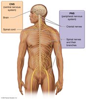
The nervous system is divided into two main structural components: the Central Nervous System (CNS) and the Peripheral Nervous System (PNS).
CNS: Consists of the brain and spinal cord. The brain contains about 100 billion neurons and is the main control center. The spinal cord contains about 100 million neurons and connects the brain to the body below the head and neck.
PNS: Composed of cranial nerves (12 pairs) and spinal nerves (31 pairs) and their branches. These nerves carry signals to and from the CNS.
Functional Divisions of the Nervous System
Sensory, Integrative, and Motor Functions
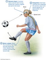
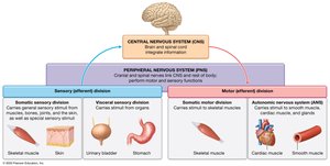
The nervous system is also divided functionally into sensory, integrative, and motor divisions.
Sensory (Afferent) Division: Gathers information from internal and external environments via sensory receptors. Includes the somatic sensory division (signals from skin, muscles, bones, joints, and special senses) and the visceral sensory division (signals from organs such as the heart, lungs, and stomach).
Integrative Functions: The CNS analyzes and interprets sensory input, determining appropriate responses. Most sensory data is filtered out as unimportant.
Motor (Efferent) Division: Executes responses by transmitting signals to effectors (muscles and glands). Includes the somatic motor division (voluntary control of skeletal muscles) and the visceral motor division (autonomic nervous system, involuntary control of smooth muscle, cardiac muscle, and glands).
Nervous Tissue
Cellular Composition
Nervous tissue is composed of about 80% cells and 20% extracellular matrix. The two main cell types are neurons and neuroglial cells.
Neurons: Excitable cells that send and receive signals (action potentials). Responsible for sensory, integrative, and motor functions.
Neuroglial Cells: Smaller, more numerous cells that support, protect, and assist neurons. They do not transmit signals.
Neurons
Structure and Function
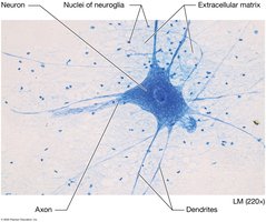
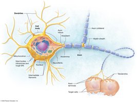
Neurons are long-lived, generally amitotic cells with three main parts: the cell body, dendrites, and axon.
Cell Body (Soma): Contains the nucleus, abundant rough endoplasmic reticulum (Nissl bodies), Golgi apparatus, nucleoli, mitochondria, and a cytoskeleton of neurofibrils and microtubules.
Dendrites: Short, branched processes that receive input and transmit electrical signals toward the cell body. Capable of growth and pruning.
Axon (Nerve Fiber): A single process that generates and conducts action potentials away from the cell body. Includes the axon hillock, axon collaterals, telodendria, and axon terminals (synaptic knobs).
Axonal Transport
Substances move along the axon via axonal transport:
Slow Axonal Transport: Moves cytoskeletal proteins away from the cell body at 1–3 mm/day.
Fast Axonal Transport: Moves vesicles and organelles using motor proteins and ATP. Can be anterograde (away from cell body, up to 400 mm/day) or retrograde (toward cell body, up to 200 mm/day).
Clinical Note: Some viruses (e.g., poliovirus, herpes simplex, rabies) and toxins (e.g., tetanus toxin) exploit retrograde axonal transport to invade the nervous system.
Functional Regions of Neurons
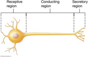
Each neuron has three main functional regions:
Receptive Region: Dendrites and cell body receive signals.
Conducting Region: Axon transmits the signal.
Secretory Region: Axon terminals secrete neurotransmitters to communicate with target cells.
Classification of Neurons
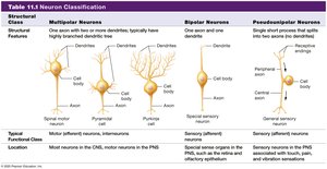
Structural Classification:
Multipolar Neurons: One axon, multiple dendrites; most common type.
Bipolar Neurons: One axon, one dendrite; found in retina and olfactory epithelium.
Pseudounipolar Neurons: Single axon with peripheral and central processes; sensory neurons for touch, pressure, and pain.
Functional Classification:
Sensory (Afferent) Neurons: Carry signals toward the CNS; usually pseudounipolar or bipolar.
Interneurons: Relay messages within the CNS; most abundant; multipolar.
Motor (Efferent) Neurons: Carry signals from the CNS to effectors; multipolar.
Structural Class | Multipolar Neurons | Bipolar Neurons | Pseudounipolar Neurons |
|---|---|---|---|
Structural Features | One axon, two or more dendrites; highly branched dendritic tree | One axon, one dendrite | Single short process splits into two axons (peripheral and central) |
Typical Functional Class | Motor (efferent) neurons, interneurons | Sensory (afferent) neurons | Sensory (afferent) neurons |
Location | Most neurons in CNS, motor neurons in PNS | Special sense organs (retina, olfactory epithelium) | General sensory neurons in PNS (touch, pain, vibration) |
Groups of Neuron Cell Bodies and Axons
Nuclei: Clusters of neuron cell bodies in the CNS.
Ganglia: Clusters of neuron cell bodies in the PNS.
Tracts: Bundles of axons in the CNS.
Nerves: Bundles of axons in the PNS.
Neuroglia (Neuroglial Cells)
Functions and Types
Neuroglia are supportive cells that maintain the environment around neurons, protect them, and assist in their function. They can divide and fill in gaps when neurons die. There are six types: four in the CNS and two in the PNS.
Astrocytes (CNS): Star-shaped cells that anchor neurons and blood vessels, facilitate nutrient and gas transport, regulate the extracellular environment, remove excess potassium and neurotransmitters, assist in forming the blood-brain barrier, and repair damaged brain tissue.
Blood-Brain Barrier and Repair
Blood-Brain Barrier: Astrocyte end-feet ensheath capillaries and induce tight junctions, selectively controlling substance exchange between blood and brain extracellular fluid.
Repair: Astrocytes divide rapidly to repair tissue but may impede neuron regrowth, sometimes causing further damage.
Clinical Application: Gliomas and Astrocytomas
Primary Brain Tumors
Gliomas are the most common primary brain tumors, originating from neuroglial cells. Astrocytomas are the most frequent type of glioma, ranging from mild to highly aggressive forms. Risk factors include ionizing radiation and genetic conditions such as neurofibromatosis. Treatment typically involves surgical removal, chemotherapy, and possibly radiation.