 Back
BackJoint Structure and Movement: Classification, Structure, and Function
Study Guide - Smart Notes
 Tailored notes based on your materials, expanded with key definitions, examples, and context.
Joint Structure and Movement
Introduction to Articulations (Joints)
Articulations, or joints, are the anatomical sites where two bones interconnect. They play a crucial role in holding bones together securely while allowing for varying degrees of mobility. The amount of movement permitted at a joint is determined by its anatomical structure.
Function of joints: Securely connect bones and allow for movement.
Mobility: Joints are the only points where bone movement occurs.
Classification of Joints
Overview of Joint Classification
Joints are classified both functionally (by the amount of movement allowed) and structurally (by their anatomical organization). Understanding these classifications is essential for identifying joint types and their roles in the body.
Functional classification: Based on range of motion (ROM).
Structural classification: Based on the material binding bones and the presence or absence of a joint cavity.
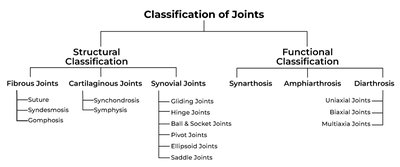
Functional Classification of Joints
Synarthrosis: Immovable joints; extremely strong (e.g., sutures in the skull).
Amphiarthrosis: Slightly movable joints; bones connected by collagen fibers or cartilage (e.g., syndesmosis, symphysis).
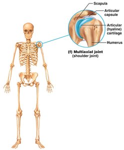
Diarthrosis: Freely movable joints; all synovial joints fall into this category.
Structural Classification of Joints
Fibrous joints: Bones joined by fibrous tissue; no joint cavity; mostly immovable.
Cartilaginous joints: Bones joined by cartilage; no joint cavity; immovable or slightly movable.
Synovial joints: Bones separated by a fluid-filled joint cavity; freely movable.
Fibrous Joints
Types of Fibrous Joints
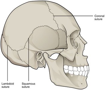
Fibrous joints are connected by dense connective tissue and generally allow little or no movement.
Suture: Found between bones of the skull; immovable.
Syndesmosis: Bones connected by a ligament; allows slight movement (e.g., between tibia and fibula).
Gomphosis: Peg-in-socket joint; found between teeth and jaws.


Cartilaginous Joints
Types of Cartilaginous Joints
Cartilaginous joints unite bones with cartilage and allow for limited movement.
Synchondrosis: Bones united by hyaline cartilage; immobile (e.g., epiphyseal plates, first rib and sternum).
Symphysis: Bones united by fibrocartilage; slightly movable (e.g., pubic symphysis, intervertebral discs).


Synovial Joints
Structure and Features of Synovial Joints
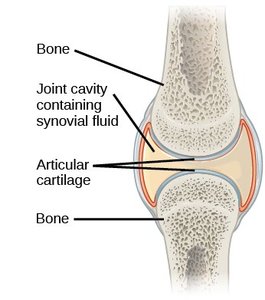
Synovial joints are the most common and movable type of joint in the body. They are characterized by a joint cavity filled with synovial fluid, which allows for a wide range of movements.
Articular cartilage: Covers the ends of bones, reducing friction and absorbing shock.
Articular capsule: Encloses the joint cavity; composed of an outer fibrous layer and an inner synovial membrane.
Joint cavity: Space containing synovial fluid.
Reinforcing ligaments: Strengthen and support the joint.
Components of Synovial Joints
Articular cartilage: Hyaline cartilage covering bone surfaces.
Joint (articular) capsule: Double-layered capsule enclosing the joint cavity.
Synovial membrane: Lines the inner surface of the capsule and secretes synovial fluid.
Synovial fluid: Lubricates, nourishes, and cushions the joint.
Ligaments: Connect bone to bone, providing stability.
Bursae and tendon sheaths: Reduce friction and facilitate movement.
Summary Table: Functional and Structural Classifications of Joints
Functional Category | Structural Category and Type | Example |
|---|---|---|
Synarthrosis (no movement) | Fibrous: Suture Fibrous: Gomphosis Cartilaginous: Synchondrosis Bony: Synostosis | Skull sutures, teeth in jaws, first rib and sternum, fused bones |
Amphiarthrosis (little movement) | Fibrous: Syndesmosis Cartilaginous: Symphysis | Tibia-fibula, pubic symphysis, intervertebral discs |
Diarthrosis (free movement) | Synovial | Shoulder, elbow, hip, knee |
Types of Synovial Joints Based on Shape
Classification by Shape and Movement
Synovial joints are further classified by the shapes of their articulating surfaces and the types of movement they allow:
Plane (gliding) joint: Allows sliding or translational movement (e.g., intercarpal joints).
Hinge joint: Permits movement in one plane (e.g., elbow).
Pivot joint: Allows rotation around a single axis (e.g., proximal radioulnar joint).
Condylar (ellipsoid) joint: Permits movement in two planes (e.g., wrist).
Saddle joint: Allows movement in two planes with greater freedom (e.g., thumb carpometacarpal joint).
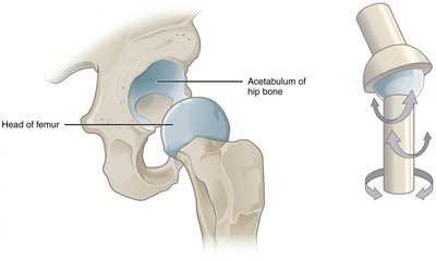
Ball-and-socket joint: Allows movement in multiple axes and planes (e.g., shoulder, hip).
Developmental and Clinical Aspects of Joints
Developmental Changes
Fetal skeletons are initially composed of hyaline cartilage and fibrous membranes.
Ossification converts these models to bone as the fetus grows.
Fontanels (soft spots) in the fetal skull allow for brain growth and skull compression during birth.
Growth of the cranium and facial skeleton continues through childhood and adolescence.
Clinical Considerations
Osteoporosis: Bone-thinning disease common in older adults, leading to increased fracture risk and vertebral collapse (kyphosis).
Dislocation (luxation): Occurs when articulating surfaces are forced out of position, damaging joint structures.
