 Back
BackJoints and Articulations: Structure, Function, and Movement
Study Guide - Smart Notes
 Tailored notes based on your materials, expanded with key definitions, examples, and context.
Joints and Articulations
Introduction
Joints, also known as articulations, are anatomical structures where two bones meet. They may be in direct contact or separated by fibrous tissue, cartilage, or fluid. Joints are essential for movement and stability in the skeletal system, and their structure determines their function and range of motion.
Classification of Joints
Joints are classified by both function and structure.
Functional Classification
- Synarthrosis: Immovable joints. Examples include sutures of the skull, teeth held in sockets, and fusion of the epiphyseal plate. 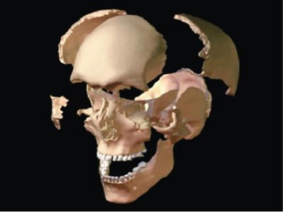 - Amphiarthrosis: Slightly movable joints. Examples include joints between radius and ulna, vertebrae, and pubic symphysis. - Diarthrosis: Freely movable joints, also known as synovial joints. These are most common in the body (arms, legs, head, etc.).
Structural Classification
- Bony fusion: Complete fusion of bones, resulting in a rigid structure (e.g., metopic suture of the frontal bone). - Fibrous joints: Bones connected by fibrous tissue. Includes sutures, syndesmoses, and gomphoses. - Cartilaginous joints: Bones connected by cartilage. Includes synchondroses and symphyses. - Synovial joints: Characterized by a joint cavity containing synovial fluid, articular cartilage, and a joint capsule.
Structure of Synovial Joints
Synovial joints are complex and allow for a wide range of movements.
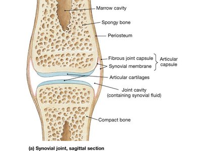
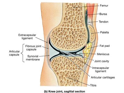
Joint capsule (articular capsule): Thick, dense connective tissue surrounding the joint.
Synovial fluid: Lubricates, nourishes chondrocytes, and acts as a shock absorber.
Articular cartilages: Hyaline cartilage covering bone surfaces.
Nerves and blood vessels: Support joint health and function.
Accessory structures: Bursae, fat pads, menisci, ligaments, and tendons stabilize and strengthen the joint.
Accessory Structures
- Bursae: Fluid-filled sacs that reduce friction between tendons/ligaments and bones. 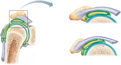 - Fat pads and menisci: Cushion and distribute pressure within the joint. - Ligaments: Connect bone to bone, providing stability. - Tendons: Connect muscle to bone, aiding movement and stability.
Types of Movements at Joints
Joints permit various types of movement, depending on their structure.
Gliding (Linear Motion)
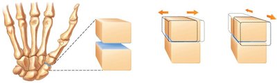
- Two opposing surfaces slide past one another. - Examples: Clavicle/sternum, carpals/metacarpals. 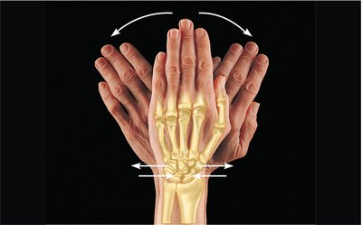
Angular Motion
- Changes the angle between bones. Includes flexion, extension, abduction, adduction, and circumduction.
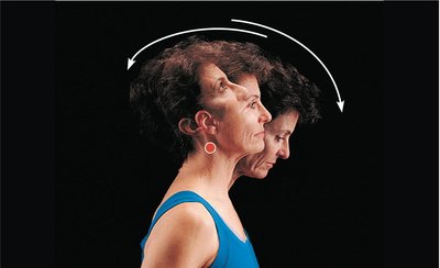
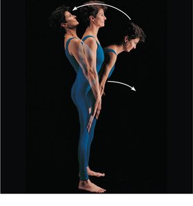
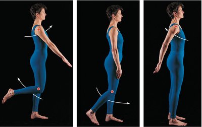
Flexion: Decreases the angle between bones.
Extension: Increases the angle between bones.
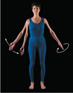
Abduction: Moves a limb away from the midline.
Adduction: Moves a limb toward the midline.
Circumduction: Circular movement combining flexion, extension, abduction, and adduction.
Rotational Movements
- Rotation occurs around a longitudinal axis. Includes head rotation, shoulder rotation, pronation, and supination. 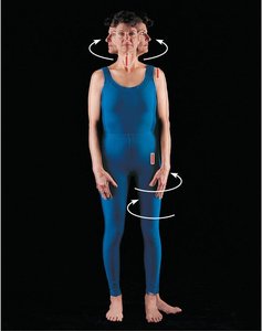
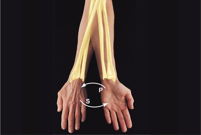
Special Movements
- Eversion/Inversion: Turning the sole of the foot laterally/medially. 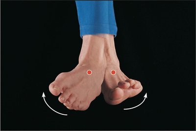 - Dorsiflexion/Plantar flexion: Lifting the foot toward the shin/depressing the foot.
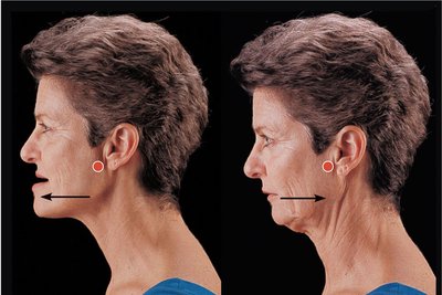
 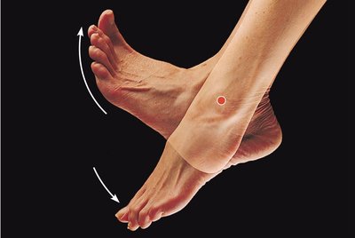 - Protraction/Retraction: Moving a body part forward/backward.
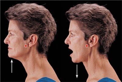
  - Elevation/Depression: Lifting/lowering a body part.
  - Opposition/Reposition: Moving the thumb to touch other fingers/returning it to anatomical position.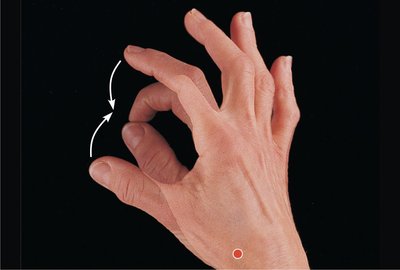
Structural Classification of Synovial Joints
Synovial joints are classified by the type and range of movement permitted.
Plane (Gliding) Joints: Allow sliding movements. Examples: ends of clavicles, between carpals/tarsals.
Hinge Joints: Permit flexion and extension in one plane. Examples: elbow, knee.
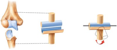
Pivot Joints: Permit rotation around a single axis. Examples: atlas/axis, radius/ulna.
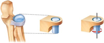
Condyloid (Ellipsoid) Joints: Permit movement in two planes (biaxial). Examples: wrist, knuckles.
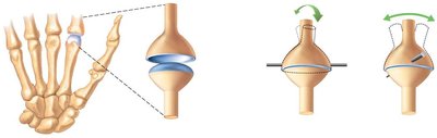
Saddle Joints: Permit biaxial movement. Example: thumb (trapezium and 1st metacarpal).
Ball-and-Socket Joints: Permit movement in multiple axes and planes (triaxial). Examples: shoulder, hip.
Major Articulations of the Axial and Appendicular Skeleton
The body contains several major joints, each with unique structural features and functions.
Temporomandibular Joint: Jaw joint, allows for complex movements including elevation, depression, protraction, and retraction.
Intervertebral Articulations: Joints between vertebrae, allow for limited movement and absorb shock.
Sternoclavicular Joint: Connects the sternum and clavicle, allows for movement of the shoulder girdle.
Glenohumeral Joint: Shoulder joint, a ball-and-socket joint with wide range of motion.
Elbow Joint: Hinge joint allowing flexion and extension of the forearm.
Wrist and Hand Joints: Complex arrangement of plane, condyloid, and saddle joints for dexterity.
Hip Joint: Ball-and-socket joint, supports body weight and allows for movement of the thigh.
Knee Joint: Largest and most complex hinge joint, includes menisci, ligaments, and bursae for stability and movement.
Ankle and Foot Joints: Permit movements such as dorsiflexion, plantar flexion, inversion, and eversion.
Joint Problems
Joints are susceptible to various disorders and injuries, including:
Arthritis: Inflammation of joints, including osteoarthritis and rheumatoid arthritis.
Meniscus Tears: Common in the knee, can impair movement and stability.
Ligament Injuries: Such as the "unhappy triad" in the knee (ruptured ACL, torn tibial collateral ligament, and torn meniscus).
Summary Table: Joint Classification
Classification | Type | Examples | Movement |
|---|---|---|---|
Functional | Synarthrosis | Skull sutures, teeth | Immovable |
Functional | Amphiarthrosis | Vertebrae, pubic symphysis | Slightly movable |
Functional | Diarthrosis | Shoulder, hip, knee | Freely movable |
Structural | Fibrous | Sutures, syndesmoses, gomphoses | Immovable/slightly movable |
Structural | Cartilaginous | Synchondroses, symphyses | Immovable/slightly movable |
Structural | Synovial | Plane, hinge, pivot, condyloid, saddle, ball-and-socket | Freely movable |
Key Equations and Concepts
- Range of Motion: Determined by joint structure, ligament strength, and muscle attachment. - Synovial Fluid Function: - Joint Stability:
Conclusion
Joints are critical for movement, stability, and overall function of the skeletal system. Understanding their classification, structure, and types of movement is essential for students of anatomy and physiology. Proper joint health is vital for mobility and quality of life.
