 Back
BackJoints and Bone Development: Structure, Function, and Regulation
Study Guide - Smart Notes
 Tailored notes based on your materials, expanded with key definitions, examples, and context.
Joints (Articulations)
Definition and Function
Joints, also known as articulations, are the sites where two or more bones meet. They are the weakest parts of the skeleton but serve essential functions:
Mobility: Joints allow movement of the skeleton.
Stability: Joints hold the skeleton together.
Classification of Joints
Structural Classification: Based on the material binding bones and the presence of a joint cavity. Types include fibrous, cartilaginous, and synovial joints.
Functional Classification: Based on movement allowed. Types include synarthroses (immovable), amphiarthroses (slightly movable), and diarthroses (freely movable).
Synovial Joints
General Structure
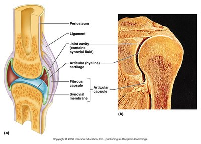
Synovial joints are characterized by the presence of a fluid-filled joint cavity and are always diarthroses (freely movable). They are found in most limb joints.
Articular cartilage: Hyaline cartilage covers the bone surfaces.
Joint (synovial) cavity: Contains synovial fluid for lubrication.
Articular capsule: Encloses the joint cavity, consisting of a fibrous capsule and synovial membrane.
Reinforcing ligaments: Strengthen and stabilize the joint.
Friction-Reducing Structures
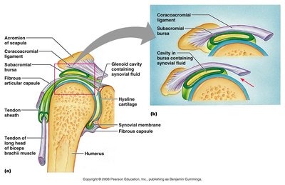
Bursae: Flattened sacs lined with synovial membrane, found where ligaments, muscles, skin, tendons, or bones rub together.
Tendon sheath: An elongated bursa that wraps around a tendon, especially in the digits.
Stability and Range of Motion
Articular surfaces: Shape determines possible movements.
Ligaments: Unite bones and prevent excessive motion.
Muscle tendons: Cross joints and help stabilize them.
Synovial joints are classified by their range of motion:
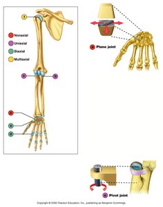
Nonaxial: Slipping movements only.
Uniaxial: Movement in one plane.
Biaxial: Movement in two planes.
Multiaxial: Movement in or around all three planes.
Types of Synovial Joints
Plane Joint
Plane joints have essentially flat articular surfaces and allow only slipping or gliding movements. They are the only examples of nonaxial joints.
Pivot Joint
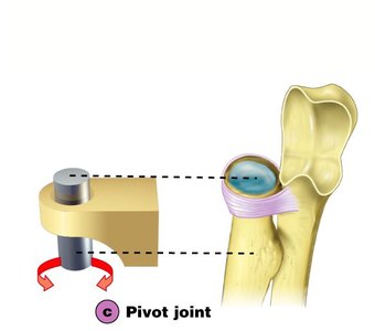
Pivot joints feature a rounded end of one bone protruding into a ring or sleeve of another bone. They allow only uniaxial movement, such as rotation. Examples include the joint between the atlas and the dens, and the proximal radioulnar joint.
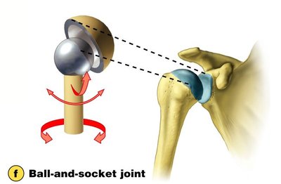
Ball-and-Socket Joint
Ball-and-socket joints have a spherical head of one bone fitting into a cuplike socket of another. These multiaxial joints permit the most freely moving synovial joints, such as the shoulder and hip.
Movements at Synovial Joints
Angular Movements
Angular movements change the angle between bones:
Flexion: Decreases the angle of the joint.
Extension: Increases the angle of the joint.
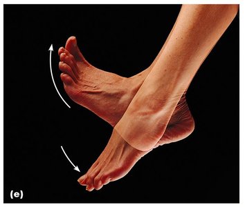
Dorsiflexion and plantar flexion: Up and down movement of the foot.
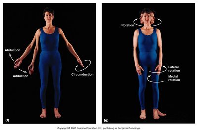
Abduction: Movement away from the midline.
Adduction: Movement toward the midline.
Circumduction: Movement describes a cone in space, combining flexion, abduction, extension, and adduction.
Rotation
Rotation is the turning of a bone around its own long axis. Examples include rotation between the first two vertebrae and at the hip and shoulder joints.
Special Movements
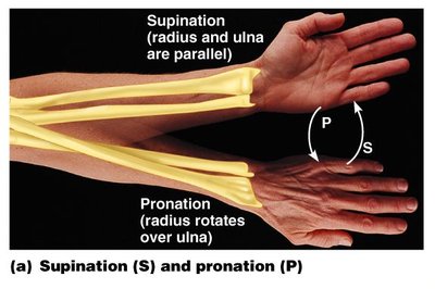
Supination and pronation: Supination is turning the palm upward; pronation is turning the palm downward.
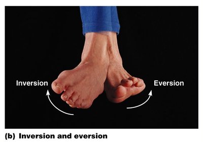
Inversion and eversion: Inversion turns the sole of the foot medially; eversion turns it laterally.
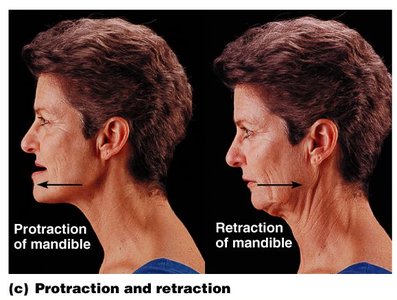
Protraction and retraction: Protraction moves a body part forward; retraction moves it backward.
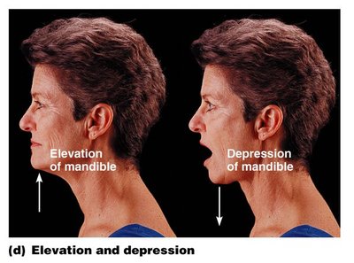
Elevation and depression: Elevation raises a body part; depression lowers it.
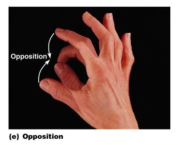
Opposition: Movement of the thumb to touch the tips of other fingers.
Bone Development and Growth
Endochondral Ossification
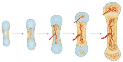
Endochondral ossification is the process by which bone forms by replacing hyaline cartilage. It begins in the second month of development and involves several stages:
Formation of bone collar around hyaline cartilage model
Cavitation of the hyaline cartilage within the cartilage model
Invasion of internal cavities by the periosteal bud and spongy bone formation
Formation of the medullary cavity; appearance of secondary ossification centers in the epiphyses
Ossification of the epiphyses; hyaline cartilage remains only in the epiphyseal plates and articular cartilages
Postnatal Bone Growth
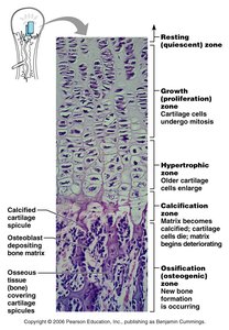
Long bones grow in length at the epiphyseal plate, which consists of several functional zones:
Resting (quiescent) zone: Relatively inactive cartilage.
Growth (proliferation) zone: Cartilage cells undergo mitosis.
Hypertrophic zone: Older cartilage cells enlarge.
Calcification zone: Matrix becomes calcified, cartilage cells die, and matrix deteriorates.
Ossification (osteogenic) zone: New bone formation occurs.
Long Bone Growth and Remodeling
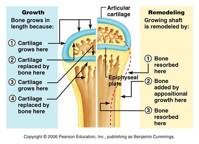
Bone grows in length as cartilage is replaced by bone at the epiphyseal plate. Remodeling occurs as bone is resorbed and added by appositional growth.
Hormonal Regulation of Bone Growth and Calcium Homeostasis
Hormonal Control of Blood Calcium
Calcium homeostasis is maintained by two hormones:
Calcitonin: Released by the thyroid gland when blood calcium levels rise. Stimulates calcium salt deposition in bone.
Parathyroid hormone (PTH): Released by the parathyroid glands when blood calcium levels fall. Stimulates osteoclasts to degrade bone matrix and release calcium into the blood.
Normal blood calcium levels are 9–11 mg/100 ml.
Summary Table: Types of Synovial Joints
Joint Type | Structure | Movement | Example |
|---|---|---|---|
Plane | Flat surfaces | Nonaxial, gliding | Intercarpal joints |
Hinge | Cylindrical projection fits into trough | Uniaxial, flexion/extension | Elbow |
Pivot | Rounded end fits into ring | Uniaxial, rotation | Proximal radioulnar joint |
Condyloid | Oval surfaces | Biaxial, angular | Wrist |
Saddle | Concave and convex surfaces | Biaxial, greater movement | Thumb |
Ball-and-Socket | Spherical head fits into socket | Multiaxial, most movement | Shoulder, hip |
Key Equations
Calcium Homeostasis:
Additional info: Academic context was added to clarify joint types, bone growth zones, and hormonal regulation mechanisms.
