 Back
BackJoints and the Skeletal System: Structure, Function, and Classification
Study Guide - Smart Notes
 Tailored notes based on your materials, expanded with key definitions, examples, and context.
Joints: Structure and Function
Definition and Importance of Joints
Joints, also known as articulations, are the locations where two or more bones meet. They are essential for movement, as bones themselves are rigid and cannot bend. The classification of joints is based on both their structural composition and their functional capacity for movement.
Joints allow movement and flexibility in the skeleton.
They are classified by structure (how bones are joined) and function (degree of movement allowed).
Structural Classification of Joints
Joints are categorized structurally into four main types:
Fibrous Joints: Bones are connected by fibrous connective tissue.
Cartilaginous Joints: Bones are connected by cartilage.
Bony Joints: Two bones fuse to form a single bone.
Synovial Joints: Bones are connected by a synovial membrane and ligaments, allowing for a wide range of movement.
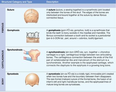
Structural Category and Type | Description |
|---|---|
Suture (Fibrous) | A synarthrotic joint found only between bones of the skull, joined by dense fibrous connective tissue. |
Gomphosis (Fibrous) | A synarthrosis that binds teeth to bony sockets in the maxillae and mandible. |
Synchondrosis (Cartilaginous) | A rigid, cartilaginous bridge between two bones, such as the connection between ribs and sternum. |
Synostosis (Bony) | An immovable joint created by the fusion of two bones, such as the epiphyseal lines of long bones. |
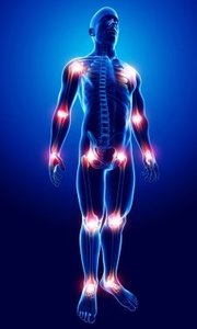
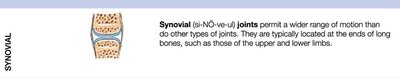
Synovial Joints
Synovial joints permit a wider range of motion than other types of joints.
They are typically located at the ends of long bones, such as those of the upper and lower limbs.
Functional Classification of Joints
Joints are also classified by their functional capacity for movement:
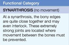
Synarthrosis: Immovable joints; extremely strong and located where movement must be prevented.
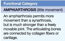
Amphiarthrosis: Slightly movable joints; stronger than freely movable joints, connected by collagen fibers or cartilage.
Diarthrosis: Freely movable joints; allow movement in one, two, or three planes.
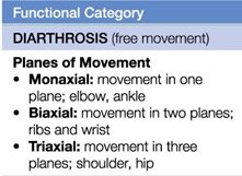
Planes of Movement in Diarthrosis
Monaxial: Movement in one plane (e.g., elbow, ankle).
Biaxial: Movement in two planes (e.g., ribs, wrist).
Triaxial: Movement in three planes (e.g., shoulder, hip).
The Axial Skeleton
Overview and Functions
The axial skeleton forms the longitudinal axis of the body and includes the bones of the head and trunk. It provides a framework that supports and protects the brain, spinal cord, and thoracic and abdominal organs.
Composed of the skull, vertebral column, and thoracic cage.
Supports and protects vital organs.
Components of the Axial Skeleton
Skull and Associated Bones: 29 bones (22 skull bones, 7 associated bones).
Thoracic Cage: 25 bones (24 ribs, 1 sternum).
Vertebral Column: 26 bones (24 vertebrae, 1 sacrum, 1 coccyx).
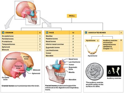
Skull: Cranial and Facial Bones
Cranial bones: Occipital, parietal, frontal, temporal, sphenoid, ethmoid.
Facial bones: Maxillae, palatine, nasal, inferior nasal conchae, zygomatic, lacrimal, vomer, mandible.
Associated bones: Hyoid bone, auditory ossicles.
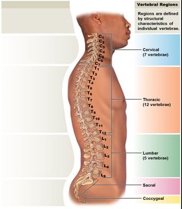
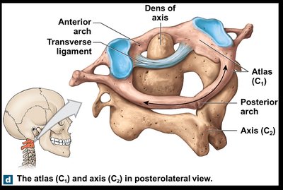
Vertebral Column
Connects to the skull and consists of 26 bones.
Divided into five regions: cervical (7), thoracic (12), lumbar (5), sacral (1), coccygeal (1).
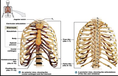
Thoracic Cage
Composed of 24 ribs and 1 sternum.
Protects organs in the thoracic cavity and allows for muscle attachment.
True ribs (1-7): Attach directly to sternum via costal cartilage.
False ribs (8-12): Not directly attached; includes vertebrochondral ribs (8-10) and floating ribs (11-12).
Sternum: Manubrium, body, xiphoid process; ossification and fusion complete by age 25.

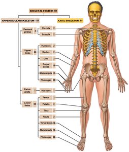
The Appendicular Skeleton
Overview and Functions
The appendicular skeleton includes the bones of the limbs and the girdles that connect them to the trunk. It is essential for movement and manipulation of the environment.
Composed of the pectoral girdle, upper limbs, pelvic girdle, and lower limbs.
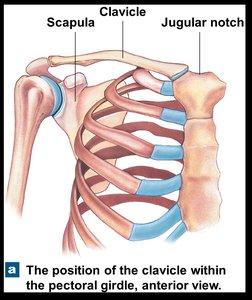
Pectoral Girdle
Attaches upper limbs to the axial skeleton.
Each girdle contains a clavicle and a scapula.
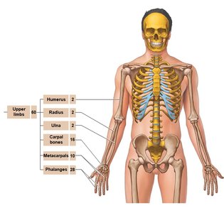
Upper Limbs
Consist of humerus (arm), radius and ulna (forearm), carpals (wrist), metacarpals and phalanges (hand).
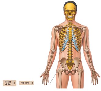
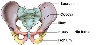
Pelvic Girdle
Attaches lower limbs to the axial skeleton.
Composed of paired pelvic bones (hip bones or coxal bones).
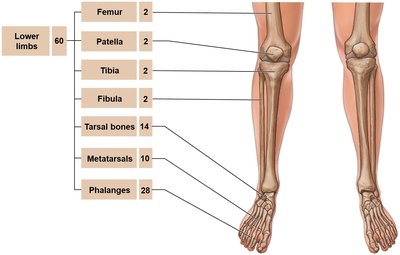
Lower Limbs
Consist of femur (thigh), patella (kneecap), tibia and fibula (leg), tarsals, metatarsals, and phalanges (foot).
Comparative Structure of Upper and Lower Limbs
The upper and lower limbs share several structural similarities:
Both have a single proximal bone (humerus in the arm, femur in the thigh).
Both have paired bones in the distal segment (radius/ulna in the forearm, tibia/fibula in the leg).
Both end in multiple small bones forming joints (carpals/tarsals, metacarpals/metatarsals, phalanges).
Take Home Points
The skeletal system is composed of bones, joints, cartilages, and ligaments.
Bones are classified according to shape and have unique bone markings.
Bone is a connective tissue with a matrix of four types of cells, collagen fibers, and a calcium phosphate ground substance.
Bone growth begins in utero and continues throughout life via bone remodeling.
Joints are classified by how bones are joined and the amount of movement allowed.
The axial skeleton consists of the skull, vertebral column, and thoracic cage.
The appendicular skeleton consists of the pectoral and pelvic girdles and the bones of the upper and lower limbs.
