 Back
BackJoints (Articulations): Structure, Classification, and Function
Study Guide - Smart Notes
 Tailored notes based on your materials, expanded with key definitions, examples, and context.
Joints (Articulations)
Definition and Overview
Joints, or articulations, are points of contact between two bones, bones and cartilage, or bones and teeth. They are essential for movement, support, and stability in the skeletal system. Joints are classified both structurally and functionally, based on their anatomical features and the degree of movement they allow.
Structural classification: Based on the presence or absence of a synovial cavity and the type of connective tissue binding the bones.
Functional classification: Based on the degree of movement permitted at the joint.
Structural Classification of Joints
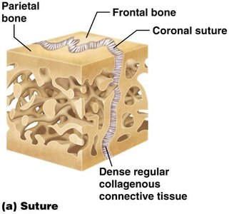
Fibrous Joints
Fibrous joints are connected by dense regular connective tissue and lack a synovial cavity. They allow little to no movement.
Sutures: Thin layer of dense regular connective tissue found between bones of the skull. Immovable (synarthrosis).
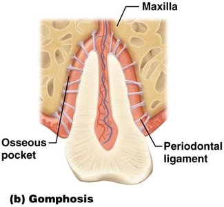
Gomphoses: Peg-in-socket joints, such as the articulation between a tooth and its socket (alveolus) in the maxilla or mandible. Slightly movable (amphiarthrosis) in children, immovable in adults.
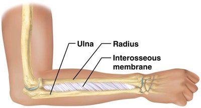
Syndesmoses: Bones connected by a sheet of dense regular connective tissue (interosseous membrane), such as between the radius and ulna. Allows slight movement (amphiarthrosis).
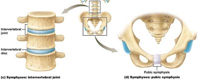
Cartilaginous Joints
Cartilaginous joints lack a synovial cavity and are united by cartilage. They allow little or no movement.
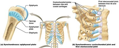
Synchondroses: Bones joined by hyaline cartilage. Examples include the epiphyseal plate in growing bones and the joint between the first rib and the sternum. Usually immovable (synarthrosis).
Symphyses: Bones joined by fibrocartilage. Examples include the pubic symphysis and intervertebral joints. Allow slight movement (amphiarthrosis).
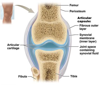
Synovial Joints
Synovial joints are characterized by the presence of a synovial cavity filled with synovial fluid. They are freely movable (diarthrosis) and are the most common type of joint in the body.
Articular cartilage: Covers the articulating surfaces of bones, reducing friction and absorbing shock.
Articular capsule: Encloses the joint cavity, consisting of an outer fibrous layer and an inner synovial membrane.
Synovial fluid: Lubricates and nourishes the joint.
Accessory structures: Menisci (pads of cartilage), bursae (fluid-filled sacs), and ligaments (extracapsular and intracapsular) provide additional support and cushioning.
Nerve supply: Provides sensory and motor information to the joint.
Functional Classification of Joints
Synarthrosis: Immovable joint (e.g., sutures of the skull).
Amphiarthrosis: Slightly movable joint (e.g., intervertebral discs, pubic symphysis).
Diarthrosis: Freely movable joint (all synovial joints).
Types of Synovial Joints
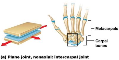
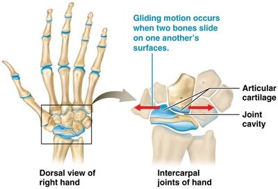
Planar (Gliding) Joints
Permit side-to-side and back-and-forth movements. Articulating surfaces are flat or slightly curved. Examples: intercarpal and intertarsal joints.
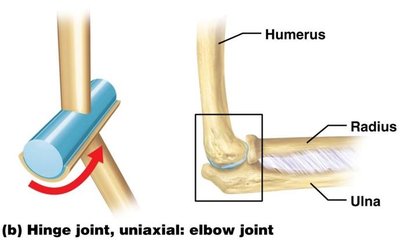
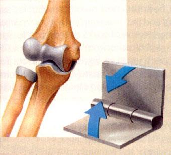
Hinge Joints
Convex surface of one bone fits into the concave surface of another, allowing movement in one plane (flexion and extension). Examples: elbow and knee joints.
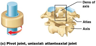
Pivot Joints
Rounded surface of one bone fits into a ring formed by another bone and ligament, allowing rotation around a single axis. Example: proximal radioulnar joint.
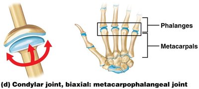
Condylar (Ellipsoid) Joints
Oval-shaped projection fits into an oval depression, allowing movement in two planes (flexion/extension, abduction/adduction). Example: metacarpophalangeal joints.
Saddle Joints
Articulating surfaces are shaped like a saddle and a rider, allowing movement in two planes. Example: carpometacarpal joint of the thumb.
Ball-and-Socket Joints
Ball-shaped surface fits into a cup-like depression, allowing movement in multiple axes and planes. Examples: shoulder and hip joints.
Types of Movements at Synovial Joints
Gliding Movements
Flat bone surfaces move back and forth or side to side with respect to one another. No significant angular or rotational movement. Example: intercarpal joints.
Angular Movements
Flexion: Decreases the angle between articulating bones.
Extension: Increases the angle between articulating bones.
Hyperextension: Extension beyond the anatomical position.
Lateral flexion: Bending the trunk to the side.
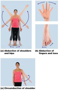
Abduction: Movement away from the midline.
Adduction: Movement toward the midline.
Circumduction: Distal end of a limb moves in a circle (combination of flexion, abduction, extension, and adduction).

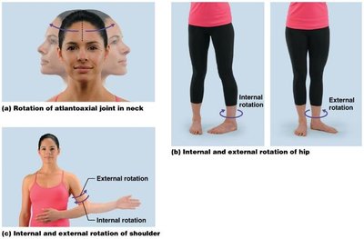
Rotational Movements
A bone revolves around its own longitudinal axis. Examples: rotation of the head, rotation of the shoulder and hip.
Special Movements
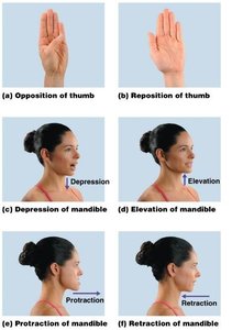
Elevation/Depression: Upward or downward movement (e.g., mandible, shoulders).
Protraction/Retraction: Anterior or posterior movement in a transverse plane (e.g., mandible, clavicle).
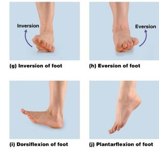
Inversion/Eversion: Movement of the sole of the foot inward or outward.
Dorsiflexion/Plantarflexion: Movement of the foot at the ankle upward or downward.
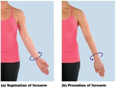
Pronation/Supination: Rotation of the forearm so the palm faces down or up.
Opposition: Movement of the thumb to touch the fingertips.
Range of Motion (ROM)
Range of motion refers to the range through which the bones of a joint can be moved. Several factors affect ROM:
Structure/shape of articulating bones
Strength and tension of ligaments
Arrangement and tension of muscles
Contact of soft parts
Hormones
Disuse
Specific Synovial Joints
Elbow Joint (Hinge Joint)
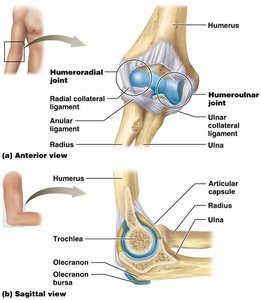
The elbow is a hinge joint formed by the trochlea of the humerus, trochlear notch of the ulna, and head of the radius. It allows flexion, extension, and rotation. Ligaments include the ulnar collateral, radial collateral, and anular ligaments.
Knee Joint (Modified Hinge Joint)
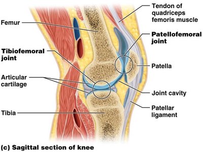
The knee is a complex joint composed of three separate joints: patellofemoral, lateral tibiofemoral, and medial tibiofemoral joints. It allows flexion and extension. Key ligaments include the anterior cruciate ligament (ACL) and posterior cruciate ligament (PCL), which limit hyperextension and sliding of the tibia.
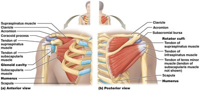
Shoulder Joint (Ball-and-Socket Joint)
The shoulder joint is formed by the head of the humerus and the glenoid cavity of the scapula. It allows rotation and almost all angular movements. The rotator cuff (tendons of four muscles) stabilizes and strengthens the joint. The articular capsule, glenoid labrum, and several ligaments provide additional support.
Hip Joint (Ball-and-Socket Joint)
The hip joint is formed by the head of the femur and the acetabulum of the pelvis. It allows rotation and almost all angular movements, with greater stability but less flexibility than the shoulder. The acetabular labrum deepens the socket, and several ligaments limit the range of motion.
Joint Replacement and Clinical Applications
Arthroscopy: Internal examination of a joint using an arthroscope.
Arthroplasty: Surgical replacement of all or part of a joint, commonly performed on the knee, hip, and shoulder.
Additional info: Joint replacement involves removing, reshaping, and replacing articulating surfaces to restore function and relieve pain.
