 Back
BackJoints (Articulations): Structure, Classification, and Function
Study Guide - Smart Notes
 Tailored notes based on your materials, expanded with key definitions, examples, and context.
Joints (Articulations)
Introduction to Joints
Joints, or articulations, are sites where two or more bones meet. They are essential for providing the skeleton with mobility and stability. Joints are classified based on their structure and the amount of movement they allow.
Articulation: The location where bones connect.
Functions of joints: Enable movement and hold the skeleton together.
Classification: Joints are classified functionally (by movement) and structurally (by binding material and cavity presence).
Classification of Joints
Functional Classification
Functional classification is based on the degree of movement permitted by the joint.
Synarthroses: Immovable joints (e.g., sutures of the skull).
Amphiarthroses: Slightly movable joints (e.g., intervertebral discs).
Diarthroses: Freely movable joints (e.g., most limb joints).
Structural Classification
Structural classification is based on the material binding the bones and the presence or absence of a joint cavity.
Fibrous joints: Bones joined by dense fibrous connective tissue; no joint cavity; mostly immovable.
Cartilaginous joints: Bones united by cartilage; no joint cavity; not highly movable.
Synovial joints: Bones separated by a fluid-filled joint cavity; all are freely movable.
Fibrous Joints
Types of Fibrous Joints
Sutures: Rigid, interlocking joints found only in the skull; allow for growth during youth and ossify in adulthood (synostoses).
Syndesmoses: Bones connected by ligaments; movement varies with fiber length (e.g., distal tibiofibular joint, interosseous membrane between radius and ulna).
Gomphoses: Peg-in-socket joints (e.g., teeth in alveolar sockets); periodontal ligament is the fibrous connection.
Cartilaginous Joints
Types of Cartilaginous Joints
Synchondroses: Bones united by hyaline cartilage (e.g., epiphyseal plates, first rib and manubrium); mostly immovable.
Symphyses: Bones united by fibrocartilage (e.g., intervertebral discs, pubic symphysis); strong, flexible, and slightly movable.
Synovial Joints
General Structure and Features
Synovial joints are the most common and movable type of joint in the body. They are characterized by the presence of a synovial cavity filled with fluid.
Articular cartilage: Hyaline cartilage covering bone ends to prevent crushing.
Joint (synovial) cavity: Small, fluid-filled space between bones.
Articular capsule: Two layers—external fibrous layer (dense irregular connective tissue) and inner synovial membrane (produces synovial fluid).
Synovial fluid: Viscous, slippery fluid that lubricates and nourishes cartilage; contains phagocytes.
Reinforcing ligaments: Capsular (thickened part of capsule), extracapsular (outside capsule), and intracapsular (deep to capsule).
Nerves and blood vessels: Nerve fibers detect pain and monitor position; capillaries supply filtrate for synovial fluid.
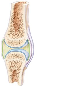
Other Features of Synovial Joints
Fatty pads: Cushioning between fibrous layer and synovial membrane or bone.
Articular discs (menisci): Fibrocartilage pads that improve fit, stabilize joint, and reduce wear.
Bursae: Sacs filled with synovial fluid that reduce friction where structures rub together.
Tendon sheaths: Elongated bursae that wrap around tendons subjected to friction.
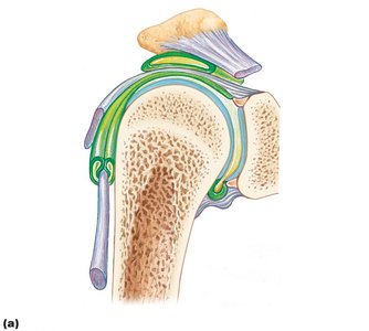
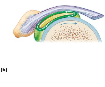
Stabilization of Synovial Joints
Factors Influencing Stability
Shape of articular surfaces: Minor role in stability.
Ligament number and location: Limited role; more ligaments usually mean more stability.
Muscle tone: Most important factor; keeps tendons taut and stabilizes joints, especially in the shoulder and knee.
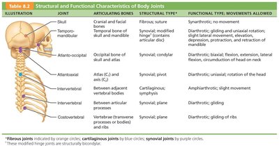
Summary Table: Structural and Functional Characteristics of Body Joints
The following tables summarize the main joints, their articulating bones, structural and functional types, and movements allowed.
Joint | Articulating Bones | Structural Type | Functional Type; Movements Allowed |
|---|---|---|---|
Skull | Cranial and facial bones | Fibrous; suture | Synarthrotic; no movement |
Temporomandibular | Temporal bone of skull and mandible | Synovial; modified hinge | Diarthrotic; gliding and uniaxial rotation |
Atlanto-occipital | Occipital bone of skull and atlas | Synovial; condyloid | Diarthrotic; biaxial, flexion, extension, lateral flexion, circumduction of head on neck |
Atlantoaxial | Atlas (C1) and axis (C2) | Synovial; pivot | Diarthrotic; uniaxial, rotation of the head |
Intervertebral | Between adjacent vertebral bodies | Cartilaginous; symphysis | Amphiarthrotic; slight movement |
Costovertebral | Vertebrae and ribs | Synovial; plane | Diarthrotic; gliding of ribs |
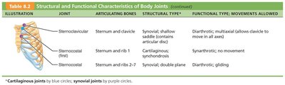
Joint | Articulating Bones | Structural Type | Functional Type; Movements Allowed |
|---|---|---|---|
Sternoclavicular | Sternum and clavicle | Synovial; shallow saddle | Diarthrotic; multiaxial |
Sternocostal (first) | Sternum and rib 1 | Cartilaginous; synchondrosis | Synarthrotic; no movement |
Sternocostal (2-7) | Sternum and ribs 2-7 | Synovial; double plane | Diarthrotic; gliding |
Joint | Articulating Bones | Structural Type | Functional Type; Movements Allowed |
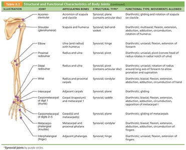
|---|---|---|---|
Shoulder (glenohumeral) | Scapula and humerus | Synovial; ball-and-socket | Diarthrotic; multiaxial, flexion, extension, abduction, adduction, rotation |
Elbow | Ulna and humerus | Synovial; hinge | Diarthrotic; uniaxial, flexion, extension |
Proximal radioulnar | Radius and ulna | Synovial; pivot | Diarthrotic; uniaxial, rotation of radius around ulna |
Wrist | Radius and carpals | Synovial; condyloid | Diarthrotic; biaxial, flexion, extension, abduction, adduction, circumduction |
Carpometacarpal (thumb) | Carpal (trapezium) and metacarpal I | Synovial; saddle | Diarthrotic; biaxial, flexion, extension, abduction, adduction, opposition |
Joint | Articulating Bones | Structural Type | Functional Type; Movements Allowed |
|---|---|---|---|
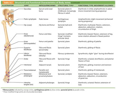
Pubic symphysis | Pubic bones | Cartilaginous; symphysis | Amphiarthrotic; slight movement |
Hip (coxal) | Hip bone and femur | Synovial; ball-and-socket | Diarthrotic; multiaxial, flexion, extension, abduction, adduction, rotation |
Knee (tibiofemoral) | Femur and tibia | Synovial; modified hinge | Diarthrotic; flexion, extension, some rotation |
Ankle | Tibia and fibula with talus | Synovial; hinge | Diarthrotic; uniaxial, dorsiflexion, plantar flexion |
Movements at Synovial Joints
Types of Movement
Gliding: Flat bone surfaces slide over each other (e.g., intercarpal joints).
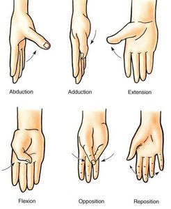
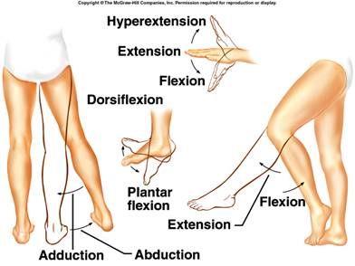
Angular movements: Change the angle between bones (flexion, extension, hyperextension, abduction, adduction, circumduction).
Rotation: Bone turns around its own long axis (e.g., rotation of the head, humerus, femur).
Special movements: Supination/pronation, dorsiflexion/plantar flexion, inversion/eversion, protraction/retraction, elevation/depression, opposition.
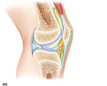
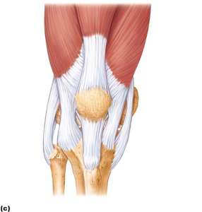
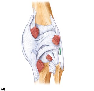
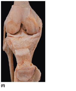
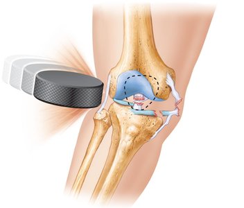
Knee Joint: Structure and Injuries
Anatomy of the Knee Joint
The knee is the largest and most complex joint in the body, consisting of three joints within a single cavity: the femoropatellar joint and the lateral and medial tibiofemoral joints. It allows flexion, extension, and some rotation.
Menisci: C-shaped fibrocartilage pads that improve fit and absorb shock.
Ligaments: Collateral, cruciate, and patellar ligaments stabilize the joint.
Bursae: At least 12 bursae reduce friction.
Knee Joint Injuries
Common injuries: Collateral ligament tears, cruciate ligament tears, and meniscal injuries (the "three C's").
Mechanism: Often caused by horizontal blows to the knee, especially when extended.
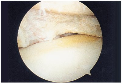
Cartilage tears: Due to compression and shear stress; cartilage heals poorly and may require arthroscopic surgery.
Sprains: Ligaments stretched or torn; partial tears heal slowly due to poor blood supply.
Dislocations (luxations): Bones forced out of alignment, often with sprains and inflammation; require reduction.
Summary
Joints are classified by structure and function, with synovial joints being the most movable and complex.
Stability is provided by articular surfaces, ligaments, and muscle tone.
Common joint injuries include sprains, dislocations, and cartilage tears, especially in the knee.
