 Back
Joints (Articulations): Structure, Classification, and Function
Study Guide - Smart Notes
 Tailored notes based on your materials, expanded with key definitions, examples, and context.
Joints (Articulations)
Introduction to Joints
Joints, also known as articulations, are anatomical structures where two or more bones meet. They play a crucial role in securing bones together and allowing varying degrees of movement throughout the body.
Definition: Joints are sites where two or more bones meet, providing both stability and mobility to the skeleton.
Classification: Joints are classified by their structure (the material binding the bones and the presence or absence of a cavity) and by their function (the amount of movement allowed).
Classification of Joints
Structural Classification
Structural classification is based on the material that binds the bones together and whether or not a joint cavity is present.
Fibrous Joints: Bones joined by dense fibrous connective tissue; no joint cavity; most are immovable.
Cartilaginous Joints: Bones joined by cartilage; no joint cavity; not highly movable.
Synovial Joints: Bones separated by a fluid-containing cavity; freely movable; most joints in the body are synovial.
Functional Classification
Functional classification is based on the amount of movement allowed by the joint.
Synarthroses: Immovable joints (e.g., sutures, tibiofibular joint, epiphyseal plate).
Amphiarthroses: Slightly movable joints (e.g., intervertebral joints, pubic symphysis).
Diarthroses: Freely movable joints; most joints in the body (e.g., shoulder, knee).
Anatomy of Joints
Fibrous Joints
Fibrous joints are united by dense fibrous connective tissue and lack a joint cavity. They are mostly immovable.
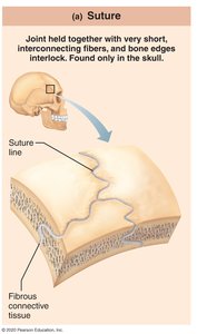
Sutures
Rigid, interlocking joints found only in the skull.
Contain short connective tissue fibers that allow for expansion in children; ossify and fuse in middle age.
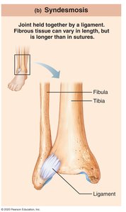
Syndesmoses
Bones connected by ligaments (bands of fibrous tissue).
Fiber length varies, so movement varies: short fibers allow little movement (e.g., tibiofibular joint), longer fibers allow more movement (e.g., radius and ulna).
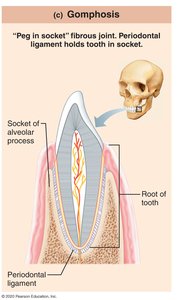
Gomphoses
Peg-in-socket joints; only example is the tooth in its alveolar socket.
The fibrous connection is the periodontal ligament, which holds the tooth in place.
Cartilaginous Joints
Cartilaginous joints are united by cartilage and lack a joint cavity. They allow little to no movement.
Synchondroses
Bar or plate of hyaline cartilage unites bones.
Almost all are synarthrotic (immovable).
Examples: epiphyseal plate, cartilage of 1st rib with manubrium of sternum.

Symphyses
Bones united by fibrocartilage; hyaline cartilage also present as articular cartilage on bony surfaces.
Strong, amphiarthrotic (slightly movable) joints.
Examples: intervertebral joints, pubic symphysis.

Synovial Joints
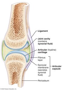
Synovial joints are characterized by a fluid-filled joint cavity that separates the articulating bones. These joints are freely movable and represent most joints in the body (e.g., shoulder, elbow, wrist, hip, knee, ankle).
Articular cartilage: Hyaline cartilage covering the ends of bones, reducing friction and absorbing shock.
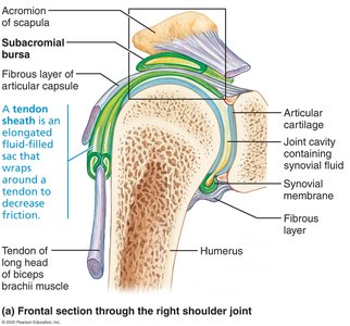
Articular (joint) capsule: Two layers thick; encloses the joint cavity. The external layer is dense irregular connective tissue (continuous with periosteum), and the inner synovial membrane is loose connective tissue that produces synovial fluid.
Synovial fluid: Viscous, slippery filtrate of plasma and hyaluronic acid; lubricates and nourishes articular cartilage and contains phagocytic cells to remove debris.

Associated Structures of Synovial Joints
Ligaments: Bands of fibrous connective tissue that reinforce and strengthen the joint.
Meniscus (Articular discs): Pads of fibrocartilage located between articular surfaces of bones; stabilize joints and reduce wear and tear (e.g., knee, sternoclavicular joint).
Bursae: Flattened sacs of fibrous connective tissue containing synovial fluid; reduce friction where ligaments, muscles, skin, tendons, or bones rub together.
Tendon sheaths: Elongated bursae that wrap completely around tendons subjected to friction.
Summary Table: Structural and Functional Classification of Joints
Structural Type | Binding Material | Joint Cavity | Functional Type | Examples |
|---|---|---|---|---|
Fibrous | Dense fibrous connective tissue | None | Synarthroses (immovable) | Sutures, syndesmoses, gomphoses |
Cartilaginous | Cartilage (hyaline or fibrocartilage) | None | Synarthroses or amphiarthroses | Synchondroses, symphyses |
Synovial | Articular cartilage, synovial fluid | Present | Diarthroses (freely movable) | Shoulder, knee, hip, elbow |
Additional Topics for Study
Factors Influencing the Stability of Synovial Joints: These include the shape of articular surfaces, the number and positioning of ligaments, and muscle tone.
Movements Allowed by Synovial Joints: Types of movement include gliding, angular movements (flexion, extension, abduction, adduction, circumduction), and rotation. Each movement type has specific examples in the body (e.g., flexion at the elbow, rotation at the atlas and axis).
Additional info: For exam preparation, students should be able to define each type of movement and provide examples, as well as describe the factors that contribute to joint stability.
