 Back
BackJoints (Articulations): Structure, Classification, and Function
Study Guide - Smart Notes
 Tailored notes based on your materials, expanded with key definitions, examples, and context.
Joints (Articulations)
Introduction to Articulations
Joints, or articulations, are the points where two or more bones meet. They serve to secure bones together and, in many cases, permit movement of the body. The structure and function of joints are essential for understanding human movement and stability.
Structural classification: Based on the type of connective tissue and the presence or absence of a joint cavity (fibrous, cartilaginous, synovial).
Functional classification: Based on the amount of movement allowed (synarthrosis, amphiarthrosis, diarthrosis).
Articulation: The point of contact between two bones or between cartilage and bone.
Key factors influencing joint movement: The fit of the bones, the type of connective tissue, and the arrangement of ligaments, muscles, and tendons.
Classification of Joints
Structural Classification
Joints are classified structurally into three main types:
Fibrous Joints: Bones are joined by dense fibrous connective tissue; no joint cavity is present. Movement is minimal or absent.
Cartilaginous Joints: Bones are united by cartilage; no joint cavity. Allow limited movement.
Synovial Joints: Bones are separated by a fluid-filled joint cavity; allow free movement.
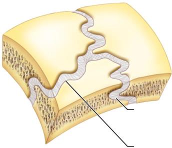
Fibrous Joints
Sutures: Interlocking joints with very short connective tissue fibers, found only in the skull. Immovable (synarthrosis), may ossify to become synostosis in adults.
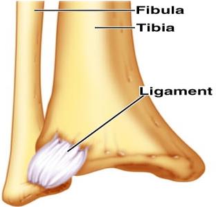
Syndesmoses: Bones connected by longer fibers (ligaments); allow slight movement (amphiarthrosis). Example: distal tibiofibular joint.
Gomphoses: Peg-in-socket joints, such as a tooth in its socket. Immovable (synarthrosis).
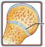
Cartilaginous Joints
Synchondroses: Bones united by hyaline cartilage; mostly immovable (synarthrosis). Examples: epiphyseal plate, first costal-sternal joint.
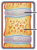
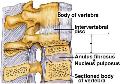
Symphyses: Bones united by fibrocartilage pad; slightly movable (amphiarthrosis). Examples: intervertebral discs, pubic symphysis.
Synovial Joints
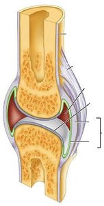
Synovial joints are the most common and freely movable joints in the body. They are characterized by the presence of a synovial cavity filled with synovial fluid.
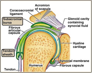
Articular cartilage: Hyaline cartilage covers the articulating bone surfaces, reducing friction and absorbing shock.
Joint (synovial) cavity: Space containing synovial fluid, which lubricates and nourishes the cartilage.
Articular capsule: Double-layered capsule enclosing the joint cavity. The outer fibrous layer provides strength; the inner synovial membrane secretes synovial fluid.
Reinforcing ligaments: Strengthen and support the joint (intrinsic, extracapsular, and intracapsular ligaments).
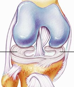
Menisci/articular discs: Pads of fibrocartilage that improve the fit between articulating bones and enhance joint stability.
Bursae, Tendon Sheaths, and Tendons
Bursae: Flattened sacs lined with synovial membrane, filled with synovial fluid. Reduce friction between moving structures (e.g., between tendon and bone).
Tendon sheaths: Elongated bursae that wrap around tendons, especially where tendons cross joints (e.g., wrist, fingers).
Tendons: Dense regular connective tissue attaching muscle to bone.
Factors Influencing the Stability of Synovial Joints
Articular surfaces: Shape and fit of the articulating surfaces (e.g., ball-and-socket joints like the hip are very stable).
Ligaments: Number and arrangement of ligaments help direct and restrict movement.
Muscle tone: The most important stabilizing factor; muscles and their tendons keep tension on the joint.
Types of Synovial Joints
Overview
Synovial joints are classified by the shape of their articular surfaces and the type of movement they allow.
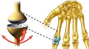
Nonaxial (Gliding/Plane) Joints
Articulating surfaces are flat or slightly curved; allow sliding or translational movements.
Examples: intercarpal and intertarsal joints.
Uniaxial Joints
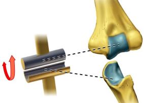
Hinge (Ginglymus) Joints: Permit flexion and extension in one plane (e.g., elbow, knee).
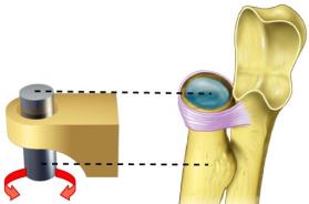
Pivot (Trochoid) Joints: Permit rotation around a single axis (e.g., proximal radioulnar joint, atlantoaxial joint).
Biaxial Joints
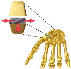
Ellipsoidal (Condyloid) Joints: Oval articular surface fits into a complementary depression; allows flexion-extension and abduction-adduction (e.g., wrist).
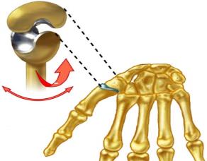
Saddle (Sellar) Joints: Each articular surface has both concave and convex areas; allows greater movement (e.g., thumb/trapezium and metacarpal).
Multiaxial Joints
Ball-and-Socket (Spheroid) Joints: Spherical head of one bone fits into a cup-like socket of another; allows movement in all axes (e.g., shoulder, hip).
Angular and Rotational Movements
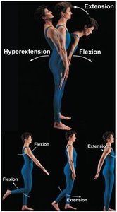
Flexion: Decreases the angle between two bones, usually in the sagittal plane.
Extension: Increases the angle between two bones; continuation beyond anatomical position is hyperextension.
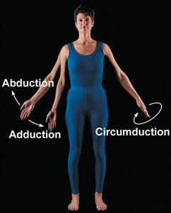
Abduction: Movement away from the midline of the body (frontal/coronal plane).
Adduction: Movement toward the midline of the body.
Circumduction: Distal end of limb moves in a circle (requires ball-and-socket joint).
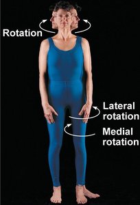
Rotation: Turning a bone around its own long axis.
Homeostatic Imbalances of Joints
Common Joint Injuries
Sprain: Stretching or tearing of ligaments without dislocation; symptoms include pain, swelling, and bruising.
Strain: Overstretching of a muscle.
Dislocation (Luxation): Bones are forced out of their normal position in the joint cavity; requires reduction.
Herniated Disc: Nucleus pulposus protrudes through the annulus fibrosus, possibly impinging on nerves.
Inflammatory and Degenerative Conditions
Bursitis: Inflammation of a bursa, often due to trauma or friction.
Tendonitis: Inflammation of tendon sheaths.
Arthritis: Over 100 types; leading cause of disability. Common forms include:
Osteoarthritis (OA): "Wear and tear" arthritis; degeneration of articular cartilage, often in large joints.
Rheumatoid Arthritis (RA): Chronic inflammatory autoimmune disorder; affects synovial membrane, leading to cartilage and bone destruction.
Gouty Arthritis (Gout): Deposition of uric acid crystals in joints, causing severe pain and inflammation, often in the big toe.
Summary Table: Classification of Joints
Structural Classification | Functional Classification | Examples |
|---|---|---|
Fibrous (sutures, syndesmoses, gomphoses) | Synarthroses (immovable), Amphiarthroses (slightly movable) | Skull sutures, distal tibiofibular joint, tooth in socket |
Cartilaginous (synchondroses, symphyses) | Synarthroses, Amphiarthroses | Epiphyseal plate, intervertebral discs, pubic symphysis |
Synovial (plane, hinge, pivot, condyloid, saddle, ball-and-socket) | Diarthroses (freely movable) | Shoulder, hip, elbow, knee, wrist, thumb |
