 Back
BackJoints (Articulations): Structure, Classification, and Movements
Study Guide - Smart Notes
 Tailored notes based on your materials, expanded with key definitions, examples, and context.
Joints (Articulations)
Definition and Functional Importance
Joints, also known as articulations, are the junctions between two or more bones. The anatomical structure of a joint determines its function and range of motion, making joints essential for movement and stability in the skeletal system.
Classification of Joints
Structural Classification
Joints are classified based on the materials that connect the bones:
Fibrous Joints: Bones are joined by dense fibrous connective tissue. These joints typically allow little or no movement.
Cartilaginous Joints: Bones are connected by cartilage, permitting limited movement.
Bony Fusion: Bones are fused together, resulting in immovable joints.
Synovial Joints: Bones are separated by a joint cavity filled with synovial fluid, allowing free movement.
Functional Classification
Synarthrosis: Immovable joints.
Amphiarthrosis: Slightly movable joints.
Diarthrosis: Freely movable joints.
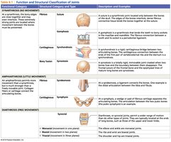
Function and Structural Classification Table
The following table summarizes the main types of joints, their structural and functional categories, and examples:
Synovial Joints
Characteristics and Components
Synovial joints are classified as diarthroses, meaning they are freely movable. They possess several specialized structures:
Fibrous Joint Capsule: Encloses the joint, providing stability.
Synovial Membrane: Produces synovial fluid for lubrication.
Articular Cartilages: Cover the bone surfaces, reducing friction.
Joint Cavity: Contains synovial fluid.
Accessory Structures: Includes menisci, fat pads, ligaments, tendons, bursae, and synovial tendon sheaths.
Sensory Nerves and Blood Vessels: Support joint function and health.
Synovial Fluid
Lubrication: Reduces friction between articular surfaces.
Nutrient Distribution: Supplies nutrients to articular cartilage.
Shock Absorption: Cushions the joint during movement.
Accessory Structures
Menisci: Fibrous cartilage pads that limit movement and stabilize the joint.
Fat Pads: Protect articular cartilages and serve as packing material.
Ligaments: Connect bone to bone, providing stability.
Tendons: Connect muscle to bone, strengthening the joint and limiting movement.
Bursae: Fluid-filled sacs that reduce friction and absorb shock.
Synovial Tendon Sheaths: Surround tendons, reducing friction.
Movements of Synovial Joints
Types of Movements
Synovial joints allow a variety of movements, including linear, angular, circumduction, rotation, and special movements at specific joints.
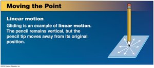
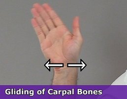
Linear Movements: Gliding
Gliding occurs when flat bone surfaces slide past each other, as seen in the carpal bones of the wrist.
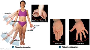
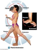
Angular Movements
Abduction: Movement away from the midline of the body.
Adduction: Movement toward the midline.
Flexion: Decreases the angle between bones (e.g., bending the elbow).
Extension: Increases the angle between bones (e.g., straightening the elbow).
Hyperextension: Extension beyond the anatomical position.
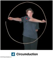
Circumduction
Circumduction is a circular movement that combines flexion, extension, abduction, and adduction, commonly seen at the shoulder and hip joints.
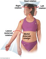
Rotational Movements
Left/Right Rotation: Rotation of the head.
Internal (Medial) Rotation: Rotation toward the midline.
External (Lateral) Rotation: Rotation away from the midline.
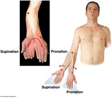
Pronation: Rotation of the forearm so the palm faces down.
Supination: Rotation of the forearm so the palm faces up.
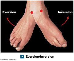
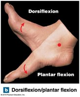
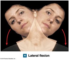
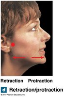
Special Movements
Eversion: Turning the sole of the foot outward.
Inversion: Turning the sole inward.
Dorsiflexion: Elevating the toes.
Plantar Flexion: Elevating the heel.
Lateral Flexion: Bending the vertebral column to the side.
Protraction: Moving a body part forward.
Retraction: Moving a body part backward.
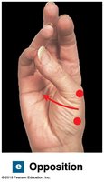
Opposition: Moving the thumb to touch the fingertips.
Reposition: Returning the thumb to its anatomical position.
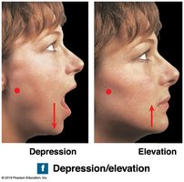
Depression: Moving a body part downward (e.g., opening the mouth).
Elevation: Moving a body part upward (e.g., closing the mouth).
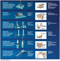
Classification of Synovial Joints
Synovial joints are further classified based on their shapes and the movements they allow:
Stability of Joints
Relationship Between Mobility and Stability
The greater the range of motion of a joint, the weaker it tends to be. Stability is enhanced by:
Ligaments and Collagen Fibers: Reinforce the joint capsule.
Shapes of Articulating Surfaces: Influence movement and stability.
Tension in Tendons: Maintains joint integrity.
Clinical Notes About Joints
Common Joint Disorders
Dislocation (Luxation): Complete displacement of a joint.
Subluxation: Incomplete or partial dislocation.
Arthritis: Inflammation of joints, leading to pain and reduced mobility.
Summary Table: Joint Types and Movements
This guide provides a comprehensive overview of joint structure, classification, and movement types, with clinical relevance for understanding joint disorders and their impact on mobility.
