Back
BackJoints (Articulations): Structure, Function, and Clinical Considerations
Study Guide - Smart Notes
 Tailored notes based on your materials, expanded with key definitions, examples, and context.
Joints (Articulations)
Introduction to Joints
Joints, or articulations, are anatomical structures where two bones connect. They are essential for movement and provide varying degrees of mobility and stability. The structure of a joint determines its function, with increased mobility often resulting in decreased strength.
Classification of Joints
Structural and Functional Classification
Joints are classified by both their anatomical structure and their functional range of motion.
Structural Classification: Based on the material binding the bones and the presence or absence of a joint cavity.
Functional Classification: Based on the amount of movement allowed at the joint.
Structural Categories of Joints
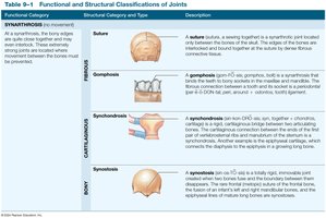
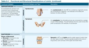
Fibrous Joints: Bones held together by dense fibrous connective tissue. Examples include sutures (skull), gomphoses (teeth in sockets), and syndesmoses (distal tibia-fibula joint).
Cartilaginous Joints: Bones joined by cartilage. Examples include synchondroses (first rib-sternum) and symphyses (pubic symphysis).
Bony Joints (Synostosis): Two bones fuse and the boundary disappears (e.g., epiphyseal lines).
Synovial Joints: Bones separated by a fluid-filled cavity, allowing free movement (e.g., knee, shoulder).
Functional Category | Structural Category and Type | Description |
|---|---|---|
Synarthrosis (Immovable) | Suture, Gomphosis, Synchondrosis, Synostosis | Immovable joints; bones may interlock or be fused. |
Amphiarthrosis (Slightly movable) | Syndesmosis, Symphysis | Joints with limited movement; bones connected by ligaments or cartilage. |
Diarthrosis (Freely movable) | Synovial | Freely movable joints with a synovial cavity. |
Synovial Joints
Basic Structure
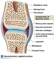
Synovial joints are diarthroses, meaning they are freely movable. They are found at the ends of long bones and are characterized by a joint capsule, synovial membrane, synovial fluid, and articular cartilage.
Joint Capsule (Articular Capsule): Encloses the joint cavity and is continuous with the periosteum of the bones.
Synovial Membrane: Lines the inner surface of the capsule and produces synovial fluid.
Synovial Fluid: Lubricates, nourishes, and absorbs shock within the joint.
Articular Cartilage: Covers bone surfaces, reducing friction and preventing bone-to-bone contact.
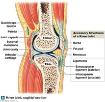
Accessory Structures
Meniscus: Fibrocartilage pad that improves fit and stability.
Fat Pads: Protect articular cartilages and fill joint spaces.
Ligaments: Strengthen and support joints; can be extracapsular or intracapsular.
Tendons: Attach muscles to bones and stabilize joints.
Bursae: Fluid-filled sacs that reduce friction and absorb shock.
Movements at Synovial Joints
Axes of Motion
Joints can be classified by the number of axes around which they permit movement:
Monaxial: Movement in one plane (e.g., elbow).
Biaxial: Movement in two planes (e.g., wrist).
Triaxial: Movement in three planes (e.g., shoulder).
]]]]]]]]]]]]]]]]]]]]]]]]]]]]]]]]]]]]]]]]]]]]]]]]]]]]]]]]]]]]]]]]]]]]]]]]]]]]]]]]]]
Types of Movement
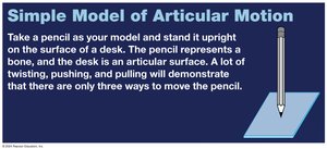
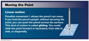
Gliding: Flat surfaces slide past each other (e.g., carpal bones).
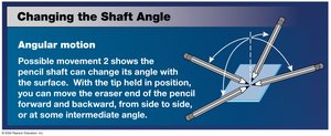
Angular Movements: Change the angle between bones.
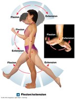
Flexion: Decreases the angle (e.g., bending elbow).
Extension: Increases the angle (e.g., straightening elbow).
Hyperextension: Extension beyond anatomical position.
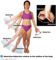
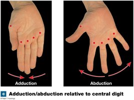
Abduction: Movement away from the midline.
Adduction: Movement toward the midline.
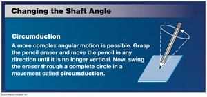
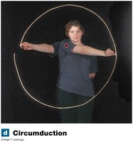
Circumduction: Circular movement without rotation.
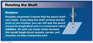
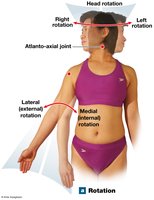
Rotational Movements: Bone turns around its own axis.
Medial (Internal) Rotation: Toward the body's midline.
Lateral (External) Rotation: Away from the body's midline.
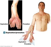
Pronation/Supination: Rotation of the forearm.
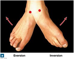
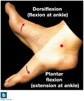
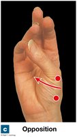
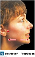
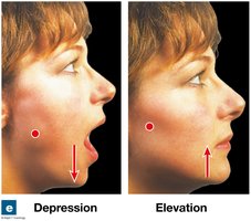
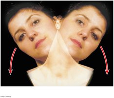
Special Movements: Unique to certain joints (e.g., inversion/eversion of the foot, dorsiflexion/plantar flexion at the ankle, opposition/reposition of the thumb, protraction/retraction, elevation/depression, lateral flexion).
Classification of Synovial Joints by Shape
Type | Movement | Example |
|---|---|---|
Plane (Gliding) | Nonaxial, gliding | Intercarpal joints |
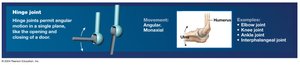
Hinge | Monaxial, angular | Elbow, knee |
Condylar (Ellipsoid) | Biaxial, angular | Radiocarpal joint |
Saddle | Biaxial, angular | First carpometacarpal joint |
Pivot | Monaxial, rotation | Atlanto-axial joint |
Ball-and-socket | Triaxial, angular, circumduction, rotation | Shoulder, hip |





Intervertebral Joints
Structure and Function
Intervertebral joints connect adjacent vertebral bodies and articular processes. The first two cervical vertebrae are joined by a pivot synovial joint, while other vertebrae are connected by plane synovial joints and symphyses.
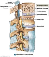
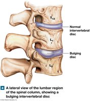
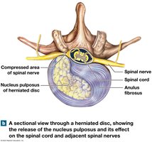
Intervertebral Discs: Fibrocartilage discs with an outer anulus fibrosus and inner nucleus pulposus, acting as shock absorbers.
Vertebral End Plates: Hyaline and fibrocartilage covering disc surfaces.
Common Injuries: Bulging disc (anulus fibrosus protrudes) and herniated disc (nucleus pulposus breaks through and compresses nerves).
Intervertebral Ligaments
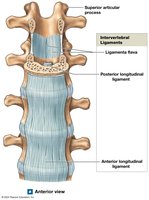
Ligamenta Flava: Connect laminae of adjacent vertebrae.
Posterior Longitudinal Ligament: Connects posterior vertebral bodies.
Anterior Longitudinal Ligament: Connects anterior vertebral bodies.
Interspinous Ligaments: Connect spinous processes.
Supraspinous Ligament: Connects tips of spinous processes, continuous with ligamentum nuchae to the skull.
Elbow and Knee Joints
Elbow Joint
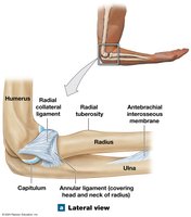
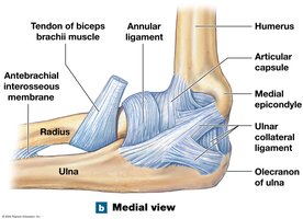
The elbow is a strong hinge joint involving the humerus, radius, and ulna. It consists of the humero-ulnar and humeroradial joints, stabilized by a thick joint capsule and strong ligaments.
Radial Collateral Ligament: Stabilizes lateral surface.
Annular Ligament: Binds head of radius to ulna.
Ulnar Collateral Ligament: Stabilizes medial surface.
Knee Joint
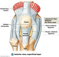
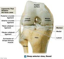
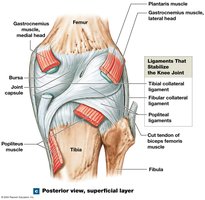
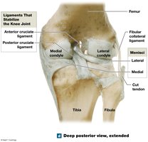
The knee is a complex hinge joint involving the femur, tibia, and patella. It transfers weight and allows flexion and extension. The joint capsule is thin and incomplete, with menisci, fat pads, and bursae for cushioning and stability.
Medial and Lateral Menisci: Fibrocartilage pads that cushion and stabilize.
Major Ligaments: Patellar, popliteal, anterior and posterior cruciate, tibial collateral, and fibular collateral ligaments.
Shoulder and Hip Joints
Shoulder Joint (Glenohumeral Joint)
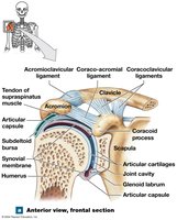
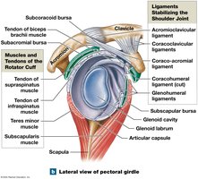
The shoulder is a ball-and-socket diarthrosis between the humerus and scapula, allowing the greatest range of motion but being the most frequently dislocated joint. Stability is provided by muscles, tendons, ligaments, and the glenoid labrum.
Ligaments: Acromioclavicular, coracoclavicular, coraco-acromial, coracohumeral, glenohumeral.
Rotator Cuff Muscles: Supraspinatus, infraspinatus, teres minor, subscapularis.
Bursae: Subdeltoid, subcoracoid, subacromial, subscapular.
Hip Joint
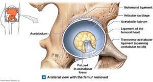
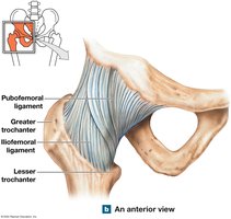
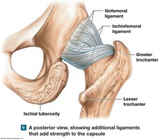
The hip is a strong ball-and-socket diarthrosis between the femur and acetabulum. It allows a wide range of movements and is stabilized by the acetabular labrum and several strong ligaments.
Ligaments: Iliofemoral, pubofemoral, ischiofemoral, transverse acetabular, ligamentum teres.
Acetabular Labrum: Deepens the socket and retains synovial fluid.
Effects of Aging on Joints
Degenerative Changes and Clinical Problems
Rheumatism: Pain and stiffness in the musculoskeletal system.
Arthritis: Inflammation of joints, including osteoarthritis (degenerative), rheumatoid arthritis (autoimmune), and gouty arthritis (uric acid crystals).
Joint Immobilization: Reduces synovial fluid flow, leading to degeneration; treated with continuous passive motion (CPM).
Increased Risk: Aging decreases bone mass and increases risk of fractures and dislocations.
Integration with Other Systems
Functional Relationships
Bone Remodeling: Involves osteoblasts (formation) and osteoclasts (resorption); influenced by age, activity, hormones, and mineral availability.
System Interactions: Muscles attach to bones; endocrine system regulates growth; skeletal system produces blood cells; digestive and urinary systems supply minerals; skeleton serves as a mineral reserve.