Back
BackLecture 4.1: Joints (Articulations)
Study Guide - Smart Notes
 Tailored notes based on your materials, expanded with key definitions, examples, and context.
Joints and Articulations
Introduction to Joints
Joints, also known as articulations, are the locations where two or more bones meet. They are essential for movement and provide mechanical support to the skeleton. The structure of a joint determines its range of motion and stability; generally, the greater the range of motion, the less stable the joint.
Joint strength and stability decrease as the range of motion increases.
Joints are classified by both their structure and function.
Classification of Joints
Functional Classification
Synarthrosis: Immovable joints (e.g., sutures of the skull).
Amphiarthrosis: Slightly movable joints (e.g., intervertebral discs).
Diarthrosis: Freely movable joints (e.g., shoulder, knee).
Structural Classification
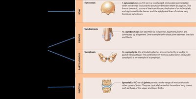
Bony: Bones fused together (e.g., synostosis).
Fibrous: Bones joined by fibrous tissue (e.g., sutures, syndesmoses).
Cartilaginous: Bones joined by cartilage (e.g., symphysis, synchondrosis).
Synovial: Bones separated by a fluid-filled cavity (e.g., most limb joints).
Structure and Types of Synovial Joints
Basic Structure of Synovial Joints
Synovial joints are the most mobile type of joint but are relatively weak. They are stabilized by accessory structures such as cartilages, fat pads, ligaments, tendons, and bursae.
Articular cartilage covers the ends of bones, reducing friction.
Synovial membrane produces synovial fluid for lubrication.
Joint capsule encloses the joint cavity.
Accessory structures include menisci, ligaments, and bursae.
Types of Synovial Joints
Synovial joints are classified by the shapes of their articulating surfaces and the types of movement they allow.
Joint Type | Movement | Examples |
|---|---|---|
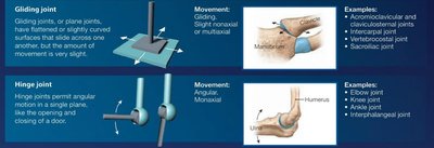
Gliding (plane) | Gliding, slight nonaxial/multiaxial | Acromioclavicular, intercarpal joints |
Hinge | Angular, monaxial | Elbow, knee, ankle joints |
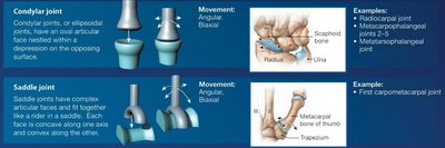
Condylar (ellipsoid) | Angular, biaxial | Radiocarpal, metacarpophalangeal joints |
Saddle | Angular, biaxial | First carpometacarpal joint (thumb) |
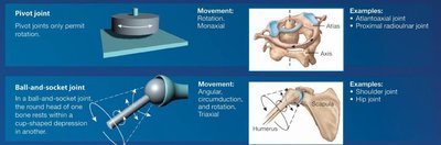
Pivot | Rotation, monaxial | Atlantoaxial, proximal radioulnar joints |
Ball-and-socket | Angular, circumduction, rotation, triaxial | Shoulder, hip joints |
Joint Motion and Axes
Simple Model of Articular Motion
Joint movement can be modeled by imagining a pencil standing upright on a desk. The pencil can move in three main ways: linear motion, angular motion, and rotation.
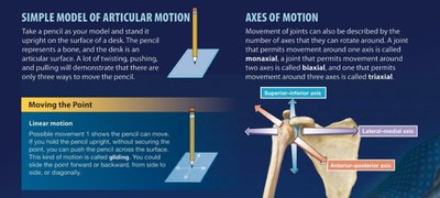
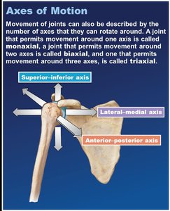
Axes of Motion
Joints can be described by the number of axes they rotate around:
Monaxial: Movement in one plane (e.g., elbow).
Biaxial: Movement in two planes (e.g., wrist).
Triaxial: Movement in three planes (e.g., shoulder).
Movements at Synovial Joints
Types of Movement
Gliding: Sliding movements between flat surfaces.
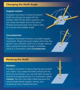
Angular: Change in angle between bones (flexion, extension, abduction, adduction).
Circumduction: Circular movement combining flexion, extension, abduction, and adduction.
Rotation: Movement around a bone's long axis (medial/lateral rotation, pronation/supination).
Special movements: Unique to certain joints (e.g., opposition, inversion, eversion).
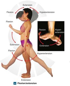
Angular Movements
Flexion: Decreases the angle between bones (e.g., bending the elbow).
Extension: Increases the angle between bones (e.g., straightening the elbow).
Hyperextension: Extension beyond the anatomical position.
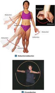
Abduction, Adduction, and Circumduction
Abduction: Movement away from the midline.
Adduction: Movement toward the midline.
Circumduction: Circular movement at a joint.
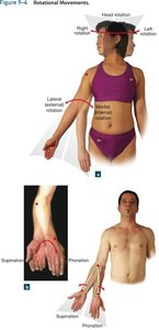
Rotational Movements
Medial (internal) rotation: Anterior surface moves toward the midline.
Lateral (external) rotation: Anterior surface moves away from the midline.
Supination: Palm faces anteriorly (upward).
Pronation: Palm faces posteriorly (downward).
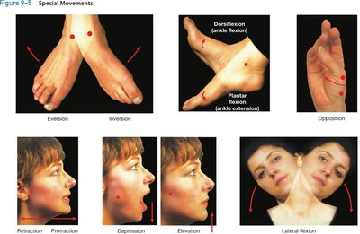
Special Movements
Eversion/Inversion: Movements of the sole of the foot outward/inward.
Dorsiflexion/Plantar flexion: Upward/downward movement of the foot at the ankle.
Opposition: Movement of the thumb to touch the fingertips.
Retraction/Protraction: Moving a part of the body backward/forward.
Depression/Elevation: Lowering/raising a body part.
Lateral flexion: Bending the vertebral column to the side.
Major Synovial Joints
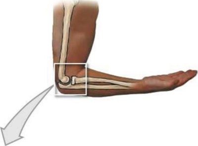
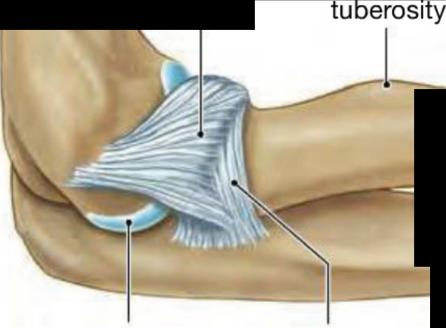
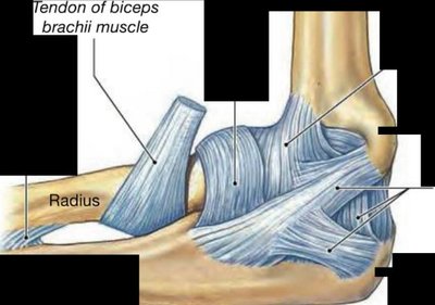
Elbow Joint
The elbow is a hinge joint formed by the humerus, radius, and ulna. It allows flexion and extension and is stabilized by strong ligaments and the bony structure of the articulating surfaces.
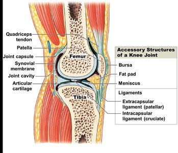
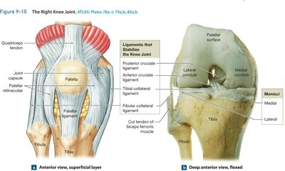
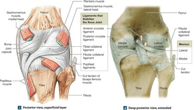
Knee Joint
The knee is a complex hinge joint that transfers weight from the femur to the tibia. It contains three articulations: two between the femur and tibia (medial and lateral condyles) and one between the patella and femur. The knee is stabilized by menisci, ligaments, and other accessory structures.
Menisci: Fibrocartilage pads that cushion and stabilize the joint.
Ligaments: Patellar, popliteal, cruciate, tibial collateral, and fibular collateral ligaments.
Shoulder and Hip Joints
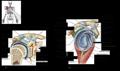
Shoulder joint: Ball-and-socket joint allowing flexion, extension, abduction, adduction, circumduction, and rotation.
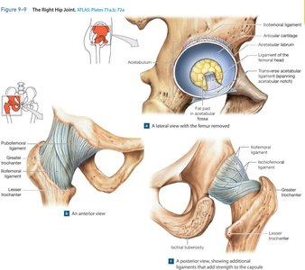
Hip joint: Ball-and-socket joint with similar movements but greater stability due to deeper socket and stronger ligaments.
Effects of Aging on Joints
Degenerative Changes
Rheumatism: Pain and stiffness in the musculoskeletal system.
Arthritis: Inflammation of joints, including osteoarthritis (wear and tear), rheumatoid arthritis (autoimmune), and gouty arthritis (uric acid crystals).
Joint immobilization reduces synovial fluid flow, increasing arthritis risk.
Aging decreases bone mass and increases fracture risk.
Integration with Other Systems
Skeletal System Interactions
Bones undergo continuous remodeling by osteoblasts (formation) and osteoclasts (recycling).
Factors affecting bone health: age, physical stress, hormones, calcium/phosphorus balance, genetics, and environment.
Muscles attach to bones for movement; endocrine, digestive, and urinary systems regulate mineral supply.
The skeleton serves as a mineral reserve and supports other organ systems.