 Back
Joints of the Body: Structure, Function, and Movements
Study Guide - Smart Notes
 Tailored notes based on your materials, expanded with key definitions, examples, and context.
Joints of the Body
Introduction to Joints
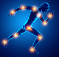
Joints, also known as articulations, are the locations where two or more bones meet. They are essential for providing movement and stability to the skeleton, allowing it to function as an integrated system. The anatomical structure of a joint determines its function, with greater mobility often resulting in reduced stability. Many common injuries, such as sprains, dislocations, and arthritis, occur at joints, making an understanding of joint anatomy crucial for prevention and treatment.
Classification of Joints
Joints are classified in two main ways: structurally and functionally. Structural classification is based on how bones are connected, while functional classification is based on the degree of movement permitted.
Structural types: Fibrous, Cartilaginous, Synovial
Functional types: Synarthrosis (immovable), Amphiarthrosis (slightly movable), Diarthrosis (freely movable)

Structural Classification of Joints
Fibrous Joints
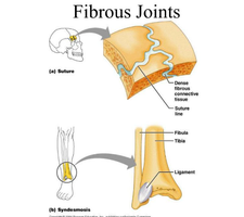
Fibrous joints are characterized by bones connected via dense connective tissue and lack a joint cavity. This absence of a cavity results in little to no movement. There are three main types:
Sutures: Found between skull bones; allow growth in children and fuse with age. Functionally classified as synarthrosis (immovable).
Syndesmoses: Bones connected by a ligament; allow limited movement. Functionally classified as amphiarthrosis (slightly movable).
Gomphosis: Peg-in-socket joint, such as a tooth in its alveolar socket. Functionally classified as synarthrosis.
Cartilaginous Joints
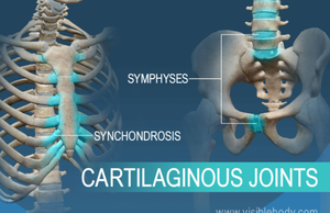
Cartilaginous joints are where bones are connected by cartilage and also lack a joint cavity. There are two main types:
Synchondrosis: Joined by hyaline cartilage (e.g., epiphyseal plate); usually immovable.
Symphysis: Bones joined by fibrocartilage (e.g., pubic symphysis); slightly movable. Symphyses absorb shock and are important in childbirth and spinal movement.
Synovial Joints
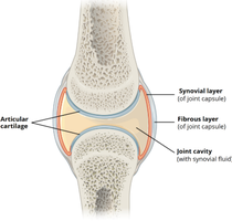
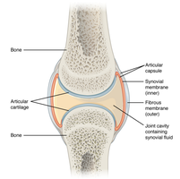
Synovial joints are the most common and most movable type of joint. They feature a joint cavity filled with synovial fluid, which lubricates, reduces friction, and nourishes cartilage. These joints are found in the limbs and permit a wide range of motion, making them common sites for injury and arthritis.
Features of Synovial Joints
General Structure
All synovial joints share several structural features:
Articular (hyaline) cartilage: Covers the ends of bones, reducing friction and absorbing shock.
Joint capsule (articular capsule): A two-layered sleeve enclosing the joint cavity.
Fibrous layer (outer): Dense connective tissue providing strength and stability.
Synovial membrane (inner): Produces synovial fluid for lubrication.
Accessory Structures
Many synovial joints include additional supportive structures:
Ligaments: Connect bone to bone, providing stability.
Tendons: Attach muscle to bone and help stabilize the joint.
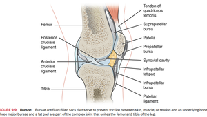
Bursa (bursae): Fluid-filled sacs that reduce friction between bone, muscle, and tendon.
Tendon sheaths: Wrap around tendons in confined spaces.
Fat pads: Provide cushioning and fill joint spaces.
Types of Synovial Joints
Classification by Shape and Movement
Synovial joints are classified based on the shape of their articulating surfaces and the movements they allow:
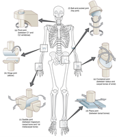
Plane (gliding) joints: Flat surfaces, allow sliding/gliding movements.
Hinge joints: Convex/concave surfaces; allow flexion and extension (elbow/knee).
Pivot joints: Rounded or pointed surface rotates within a ring; allow rotation.
Condyloid (ellipsoidal) joints: Oval condyle fits into elliptical cavity; allow flexion, extension, abduction, adduction.
Saddle joints: Concave surface fits into complementary surface; allow similar movement to condyloid joints (thumb).
Ball-and-socket joints: Spherical heads fit into cup-shaped socket; allow movement in all axes and rotation (shoulder/hip).
Movements Allowed at Joints
Gliding and Rotational Movements
Synovial joints allow various types of movement depending on their structure:
Gliding movements: Flat bone surfaces glide past one another; limited range; typical of plane joints (intercarpal, intertarsal, vertebral articular process joints).
Rotational movements: A bone turns around its own longitudinal axis.
Medial rotation: Turns towards the body’s midline.
Lateral rotation: Turns away from the midline.
Examples: Rotation of the head, shoulder, and hip joints.
Angular Movements
Angular movements change the angle between bones at a joint:
Flexion: Decreases the angle (bending elbow/knee).
Extension: Increases the angle (straightening elbow/knee).
Hyperextension: Movement beyond anatomical position.
Abduction: Movement away from midline.
Adduction: Movement toward the midline.
Circumduction: Circular movement combining flexion, extension, abduction, and adduction.
Special Movements
Special movements occur only at specific joints:
Elevation and depression: Lifting/lowering a body part (shoulders/mandible).
Protraction and retraction: Moving anteriorly/posteriorly (jaw, scapula).
Supination and pronation: Turning palm up/downward (forearm).
Dorsiflexion and plantar flexion: Flexing/pointing the foot.
Inversion and eversion: Turning the sole of the foot inward/outward.
Opposition and reposition: Thumb movement toward/away from fingers.
Anatomy of Selective Synovial Joints
Vertebral Column Joints
The vertebral column contains both symphysis and synovial plane joints:
Intervertebral joints: Symphysis joints formed by intervertebral discs (fibrocartilage); cushion and absorb shock.
Synovial plane joints: Between articular processes (zygapophyseal/facet joints); allow limited gliding movement.
Movements: Flexion, extension, lateral flexion, rotation.
Specialized cervical joints:
Atlanto-occipital joint (C1-Skull): Synovial condyloid joint; permits flexion and extension ('yes' motion).
Atlantoaxial joint (C1-C2): Synovial pivot joint; allows rotation ('no' motion).
Temporomandibular Joint (TMJ)
The TMJ allows for opening and closing of the mouth and is a combination of hinge and gliding/plane synovial joints. Movements include elevation, depression, protraction, retraction, and slight rotation. The joint is supported by several ligaments and an articular disc divides the cavity into two chambers.
Shoulder (Glenohumeral) Joint
The shoulder joint is a ball-and-socket synovial joint formed by the head of the humerus and the glenoid cavity of the scapula. It allows for a wide range of movements and is stabilized by rotator cuff muscles, ligaments, and bursae.
Elbow Joint
The elbow joint consists of two articulations within one capsule: the humeroulnar (main hinge movement) and humeroradial joints. It is a hinge joint allowing flexion and extension, stabilized by collateral ligaments and controlled by tendons of the biceps and triceps.
Hip Joint
The hip is a ball-and-socket joint formed by the head of the femur and the acetabulum. It allows for flexion, extension, abduction, adduction, rotation, and circumduction. Strong ligaments and the acetabular labrum provide stability, and bursae cushion tendons.
Knee Joint
The knee joint consists of three articulations within a single capsule: patellofemoral, lateral tibiofemoral, and medial tibiofemoral joints. It is a synovial hinge joint with slight rotation, stabilized by menisci, cruciate and collateral ligaments, and bursae.
Ankle and Foot Joints
The ankle (talocrural) joint is a hinge joint allowing dorsiflexion and plantar flexion, stabilized by medial and lateral ligaments. The subtalar joint is a gliding joint allowing inversion and eversion. Tarsal and metatarsal joints permit gliding movements, and phalangeal joints allow flexion, extension, abduction, and adduction of toes. Tendons and arches support weight and mobility.
Joint Development and Aging
Embryonic Development and Changes with Age
Joints form during embryonic development alongside bones. Mesenchyme differentiates into joint structures where bones meet. Fibrous joints form when dense connective tissue remains between developing bones, cartilaginous joints form when cartilage persists, and synovial joints form when a cavity develops and fills with synovial fluid. At birth, joints are fully formed but continue to strengthen with movement and growth. Aging decreases joint flexibility, thins cartilage, reduces synovial fluid, and increases stiffness.
Summary Table: Joint Classification and Features
Structural Type | Functional Type | Examples | Movement |
|---|---|---|---|
Fibrous | Synarthrosis, Amphiarthrosis | Sutures, Syndesmoses, Gomphosis | Immovable or slightly movable |
Cartilaginous | Synarthrosis, Amphiarthrosis | Synchondrosis, Symphysis | Immovable or slightly movable |
Synovial | Diarthrosis | Shoulder, Elbow, Hip, Knee, Ankle | Freely movable |
Key Equations and Concepts
Joint Movement Angles
Angular movement at joints can be described mathematically:
Flexion/Extension: Change in angle between bones, measured in degrees.
For example, the angle at the elbow during flexion:
where is the change in angle.
Additional info:
Rotator cuff muscles include supraspinatus, infraspinatus, teres minor, and subscapularis.
Menisci in the knee are fibrocartilaginous structures that help distribute weight and reduce friction.
Synovial fluid is produced by the synovial membrane and contains hyaluronic acid for lubrication.
