 Back
Joints of the Lower Limb: Structure, Function, and Clinical Applications
Study Guide - Smart Notes
 Tailored notes based on your materials, expanded with key definitions, examples, and context.
Joints of the Lower Limb
Hip Joint
The hip joint is a ball-and-socket synovial joint formed between the head of the femur and the acetabulum of the pelvis. It is designed for stability and weight-bearing, allowing a wide range of motion while supporting the body's weight during standing, walking, and running.
Ligaments of the Hip: The hip joint is reinforced by several strong ligaments that stabilize the joint and limit excessive movement.
Iliofemoral Ligament: Located anteriorly, this is one of the strongest ligaments in the body and prevents hyperextension of the hip.
Pubofemoral Ligament: Found anteroinferiorly, it limits excessive abduction and extension.
Ischiofemoral Ligament: Located posteriorly, it stabilizes the hip and limits internal rotation.
Ligamentum Teres (Ligament of the Head of Femur): This ligament extends from the acetabulum to the fovea capitis of the femur and adds strength to the femoral head, though it is not a major stabilizer in adults.
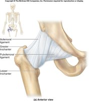
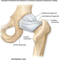
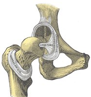
Example: The iliofemoral ligament is crucial in preventing the trunk from falling backward during standing.
Hip Replacement
Hip replacement, or total hip arthroplasty, is a surgical procedure in which a damaged hip joint is replaced with artificial components. This procedure is commonly performed to relieve pain and restore function in patients with severe arthritis or hip fractures.
Components: The artificial hip typically consists of a metal or ceramic femoral head, a metal stem, and a polyethylene or ceramic acetabular cup.
Indications: Severe osteoarthritis, rheumatoid arthritis, avascular necrosis, or traumatic injury.
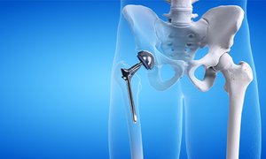
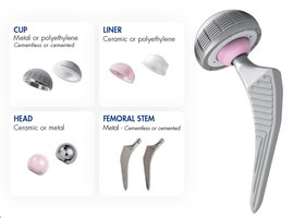
Example: A patient with advanced osteoarthritis who experiences chronic hip pain and limited mobility may benefit from hip replacement surgery.
Knee Joint
Anatomy of the Knee Joint
The knee joint is a complex hinge joint formed by the articulation of the femur, tibia, and patella. It is stabilized by multiple ligaments, tendons, and menisci, allowing flexion, extension, and slight rotation.
Major Ligaments: Anterior cruciate ligament (ACL), posterior cruciate ligament (PCL), medial collateral ligament (MCL), and lateral collateral ligament (LCL).
Menisci: Medial and lateral menisci are fibrocartilaginous structures that cushion and stabilize the joint.
Bursae: Fluid-filled sacs that reduce friction between tissues around the knee. Examples include the prepatellar, suprapatellar, and infrapatellar bursae.

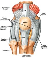

Example: The ACL prevents anterior displacement of the tibia relative to the femur.
Knee Injuries and Replacement
Knee injuries are common in athletes and the general population, with ligament tears (especially ACL) and meniscal injuries being frequent. Knee replacement (arthroplasty) is performed to restore function in severely damaged joints.
ACL Tear: Often occurs due to sudden changes in direction or impact, leading to instability.
Knee Replacement: Involves replacing damaged joint surfaces with artificial components. Can be unicompartmental (one part) or bicompartmental (multiple parts).

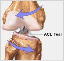

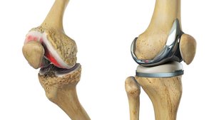
Example: An ACL tear is common in sports like football and may require surgical reconstruction.
Ankle Joint and Foot
Ankle Joint Structure and Ligaments
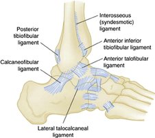
The ankle joint is a hinge-type synovial joint formed by the tibia, fibula, and talus. It is stabilized by several ligaments that prevent excessive inversion and eversion.
Major Ligaments: Anterior talofibular, posterior talofibular, calcaneofibular, and deltoid ligaments.
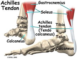
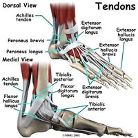
Tendons: The Achilles tendon (calcaneal tendon) is the strongest tendon in the body, connecting the calf muscles to the calcaneus.

Common Ankle and Foot Conditions
Several conditions can affect the ankle and foot, including sprains, plantar fasciitis, and tendon injuries.
Ankle Sprain: Injury to the ligaments of the ankle, graded I-III based on severity.
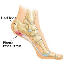
Plantar Fasciitis: Inflammation of the plantar fascia, causing heel pain.

Example: A Grade II ankle sprain involves partial tearing of the ligament and moderate swelling.
Summary Table: Major Ligaments of the Lower Limb Joints
Joint | Major Ligaments | Function |
|---|---|---|
Hip | Iliofemoral, Pubofemoral, Ischiofemoral, Ligamentum Teres | Stabilize hip, limit excessive movement |
Knee | ACL, PCL, MCL, LCL, Menisci | Stabilize knee, prevent abnormal motion |
Ankle | Anterior/Posterior Talofibular, Calcaneofibular, Deltoid | Prevent excessive inversion/eversion |
Additional info: The notes above include expanded academic context on joint structure, function, and clinical relevance, as well as examples and a summary table for clarity.
