 Back
BackJoints: Structure, Classification, and Function
Study Guide - Smart Notes
 Tailored notes based on your materials, expanded with key definitions, examples, and context.
Joints: Structure, Classification, and Function
I. Classification of Joints
Joints, or articulations, are sites where two or more bones meet. They are classified both structurally and functionally, which helps in understanding their anatomy and the range of movements they allow.
Structural Classification: Based on the material binding the bones and the presence or absence of a joint cavity. The three main types are:
Fibrous joints
Cartilaginous joints
Synovial joints
Functional Classification: Based on the amount of movement permitted:
Synarthroses: Immovable joints
Amphiarthroses: Slightly movable joints
Diarthroses: Freely movable joints
II. Fibrous Joints
Fibrous joints are connected by dense fibrous connective tissue and lack a joint cavity, resulting in little to no movement.
Sutures: Found between skull bones, held together by short connective tissue fibers.
Syndesmoses: Bones connected by a ligament (e.g., distal tibiofibular joint).
Gomphoses: Peg-in-socket joints (e.g., teeth in alveolar sockets).
III. Cartilaginous Joints
Cartilaginous joints unite bones with cartilage and lack a joint cavity, allowing limited movement.
Synchondroses: Bones joined by hyaline cartilage (e.g., epiphyseal plates in children).
Symphyses: Bones joined by fibrocartilage (e.g., pubic symphysis, intervertebral discs).
IV. Synovial Joints
Synovial joints are the most movable type of joint in the body, characterized by a fluid-filled joint cavity.
Articular cartilage covers the ends of bones, reducing friction.
Joint (synovial) cavity contains synovial fluid for lubrication.
Articular capsule encloses the joint cavity, consisting of a fibrous layer and a synovial membrane.
Synovial fluid lubricates and nourishes the articular cartilage.
Reinforcing ligaments strengthen the joint.
Nerves and blood vessels supply the joint, detecting pain and stretch, and providing nutrients.
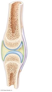
Bursae and tendon sheaths reduce friction where ligaments, muscles, skin, tendons, or bones rub together.
Factors Influencing Synovial Joint Stability
Articular surface shape determines possible movements but minimally affects stability.
Ligaments prevent excessive movement and stabilize the joint.
Muscle tone is the most important stabilizing factor, keeping tendons taut.
Movements Allowed by Synovial Joints
Gliding: Flat bone surfaces slide past each other.
Angular movements: Change the angle between bones.
Flexion: Decreases joint angle.
Extension: Increases joint angle.
Dorsiflexion/Plantar flexion: Movements at the ankle joint.
Abduction/Adduction: Movement away from/toward the midline.
Circumduction: Limb describes a cone in space.
Rotation: Bone turns around its own long axis.
Special movements: Supination, pronation, inversion, eversion, protraction, retraction, elevation, depression, opposition.
Types of Synovial Joints
Plane: Flat surfaces, gliding movements.
Hinge: Cylindrical projection fits into a trough, movement in one plane (e.g., elbow).
Pivot: Rounded end fits into a ring, rotation (e.g., proximal radioulnar joint).
Condylar: Oval surface fits into depression, all angular movements (e.g., wrist).
Saddle: Each surface has concave and convex areas, more movement than condylar (e.g., thumb).
Ball-and-socket: Spherical head fits into a socket, multiaxial movement (e.g., shoulder, hip).
V. Selected Synovial Joints
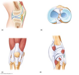
Knee Joint
The knee is the largest and most complex joint, consisting of three joints in one: femoropatellar, lateral, and medial tibiofemoral joints. It is stabilized by ligaments, menisci, and muscle tendons.
Femoropatellar joint: Plane joint, allows patella to glide.
Tibiofemoral joint: Hinge joint, allows flexion/extension and slight rotation.
Ligaments: Capsular, extracapsular, and intracapsular (cruciate) ligaments stabilize the joint.
Muscle tendons: Quadriceps and semimembranosus tendons reinforce the capsule.
Shoulder (Glenohumeral) Joint
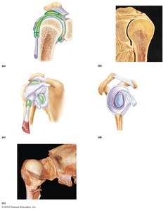
The shoulder is a ball-and-socket joint, providing the greatest range of motion but less stability. The rotator cuff muscles and tendons are the main stabilizers.
Joint capsule: Thin and loose, with weak supporting ligaments.
Rotator cuff: Group of tendons and muscles encircling the joint.
Elbow Joint
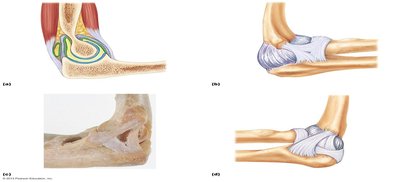
The elbow is a hinge joint between the humerus and ulna, allowing flexion and extension. The radius rotates for pronation and supination.
Articular capsule: Thin, allows movement but is stabilized by strong ligaments.
Biceps and triceps tendons: Provide additional stability.
Radius: Rotates within the annular ligament for forearm rotation.
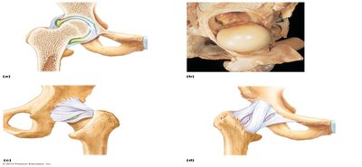
Hip (Coxal) Joint
The hip is a ball-and-socket joint, providing a wide range of motion and high stability due to the deep socket and strong ligaments.
Ligaments: Reinforce the capsule and stabilize the joint.
Muscle tendons: Contribute to stability, but the deep acetabulum is the main stabilizer.
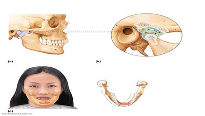
Temporomandibular Joint (TMJ)
The TMJ allows both hingelike and side-to-side movements. It contains an articular disc dividing the cavity and is reinforced by a lateral ligament.
Articular disc: Divides the synovial cavity for different movements.
Lateral ligament: Reinforces the joint.
VI. Homeostatic Imbalances of Joints
Common Joint Injuries
Sprains: Stretching or tearing of ligaments; may heal or require surgery.
Dislocations: Bones forced out of alignment, often with sprains and inflammation.
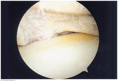
Cartilage tears: Common in the knee, due to compression and shear stress.
Inflammatory and Degenerative Conditions
Bursitis: Inflammation of a bursa, usually from friction or trauma.
Tendonitis: Inflammation of a tendon, often from overuse.
Arthritis: General term for joint inflammation and degeneration.
Osteoarthritis: Degeneration of articular cartilage and bone thickening.
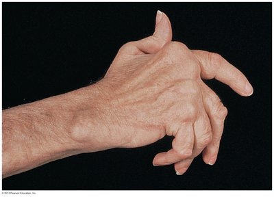
Rheumatoid arthritis: Autoimmune, chronic inflammatory disorder.
Gouty arthritis: Uric acid crystal deposition in joints.
Lyme disease: Inflammatory joint condition caused by tick-borne bacteria.
VII. Developmental Aspects of Joints
Embryonic development: Joints form alongside bones, resembling adult joints by week 8.
Childhood: Joint use shapes size, flexibility, and function.
Aging: Ligaments and tendons shorten and weaken, discs may herniate, and osteoarthritis often develops.
