 Back
BackJoints: Structure, Classification, and Function
Study Guide - Smart Notes
 Tailored notes based on your materials, expanded with key definitions, examples, and context.
Joints: Classification and Function
Overview of Joints
Joints, also known as articulations, are the sites where two or more bones meet. They provide mobility to the skeleton and maintain the integrity of the skeletal structure. Joints are classified based on their structure and function.
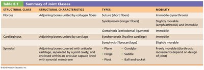
Structural classification: Based on the material binding the bones and the presence or absence of a joint cavity. Types include fibrous, cartilaginous, and synovial joints.
Functional classification: Based on the degree of movement allowed. Types include synarthroses (immovable), amphiarthroses (slightly movable), and diarthroses (freely movable).
Structural Classes of Joints
Fibrous Joints: Bones are joined by dense connective tissue and lack a joint cavity. Most are immovable. Types include sutures, syndesmoses, and gomphoses.
Cartilaginous Joints: Bones are united by cartilage and lack a joint cavity. They are not highly movable. Types include synchondroses and symphyses.
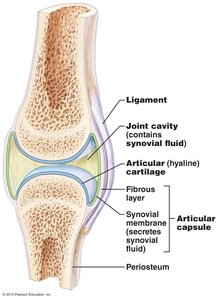
Synovial Joints: Most joints in the body. They are highly movable and have a fluid-filled joint cavity. Distinguished by six features: articular cartilage, articular cavity, articular capsule, synovial fluid, reinforcing ligaments, and nerves/blood vessels.
Functional Classes of Joints
Synarthroses: Immovable joints (e.g., sutures in the skull).
Amphiarthroses: Slightly movable joints (e.g., syndesmoses, symphyses).
Diarthroses: Freely movable joints (e.g., synovial joints).
Fibrous Joints
Sutures
Sutures are joints held together by very short, interconnecting fibers, and bone edges interlock. Found only in the skull, they are immovable and provide protection for the brain. 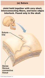
Syndesmoses
Syndesmoses are joints held together by ligaments. The fibrous tissue can vary in length but is longer than in sutures. These joints allow slight movement, such as the distal tibiofibular joint. 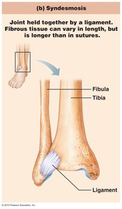
Gomphoses
Gomphoses are "peg-in-socket" fibrous joints. The periodontal ligament holds the tooth in its socket within the alveolar process of the jaw. 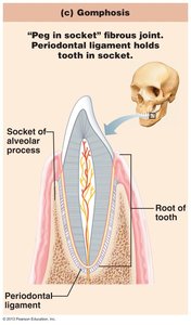
Cartilaginous Joints
Synchondroses
Synchondroses are joints where bones are united by hyaline cartilage. Most are immovable, such as the epiphyseal plate in growing bones and the joint between the first rib and sternum. 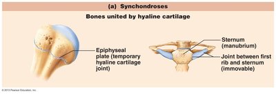
Symphyses
Symphyses are joints where bones are united by fibrocartilage. These joints are slightly movable and provide strength and flexibility, such as the pubic symphysis and intervertebral discs. 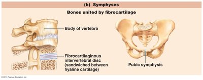
Synovial Joints
General Structure
Synovial joints are characterized by a fluid-filled joint cavity and are the most movable type of joint. They have six distinguishing features:
Articular Cartilage: Hyaline cartilage covers the ends of bones, reducing friction and absorbing shock.
Articular Cavity: Space filled with synovial fluid.
Articular Capsule: Two-layered membrane; the outer fibrous layer is dense irregular connective tissue, and the inner synovial membrane is loose connective tissue that produces synovial fluid.
Synovial Fluid: Lubricates the joint, nourishes cartilage, and reduces friction.
Reinforcing Ligaments: Strengthen and stabilize the joint.
Nerves and Blood Vessels: Provide sensory information and nutrients.
Bursae and Tendon Sheaths
Bursae and tendon sheaths are small sacs filled with synovial fluid that reduce friction between moving surfaces. 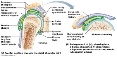
Types of Synovial Joints
Synovial joints are classified by the shape of their articulating surfaces and the movements they allow:
Plane: Flat articular surfaces; nonaxial movement (e.g., intercarpal joints).
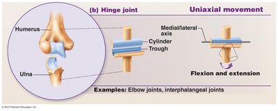
Hinge: Cylinder fits into a trough; uniaxial movement (e.g., elbow, knee).
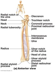
Pivot: Rounded surface fits into a ring; uniaxial movement (e.g., proximal radioulnar joint).
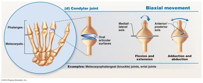
Condylar: Oval articular surfaces; biaxial movement (e.g., metacarpophalangeal joints).
Saddle: Each articular surface has both concave and convex areas; biaxial movement (e.g., thumb carpometacarpal joint).
Ball and Socket: Spherical head fits into a cup; multiaxial movement (e.g., shoulder, hip).
Joint Stability
Joint stability is influenced by:
Articular Surface: Shape determines movement and stability; deep sockets are more stable than shallow ones.
Ligaments: Unite bones and prevent excessive movement; more ligaments increase stability.
Muscle Tone: Muscle tendons crossing joints are the most important stabilizers; continuous slight contraction keeps tendons taut.
Movements at Synovial Joints
Types of Movements
Movements are classified as gliding, angular, and rotational.
Gliding: Flat bone surfaces slide over each other.
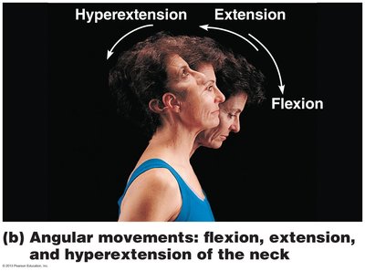
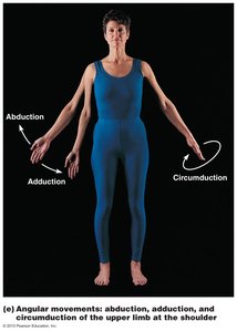
Angular: Change the angle between bones. Includes flexion, extension, hyperextension, abduction, adduction, and circumduction.
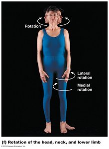
Rotation: Bone turns around its long axis; includes medial and lateral rotation.
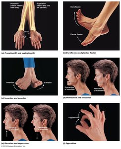
Special Movements: Include supination/pronation, dorsiflexion/plantarflexion, inversion/eversion, protraction/retraction, elevation/depression, and opposition.
Major Synovial Joints
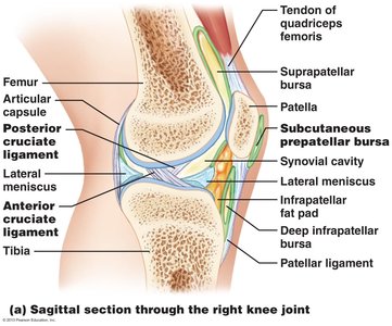
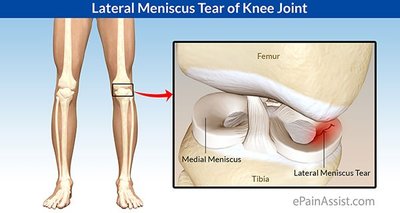
Knee Joint
The knee is the most complex joint, capable of withstanding compression, flexion, tension, and side-to-side movements. It consists of three joints in one cavity: tibiofemoral (medial and lateral meniscus), femoropatellar, and popliteus.
Articular capsule: Surrounds posterior, lateral, and medial surfaces.
Ligaments: Patellar, medial/lateral patellar retinacula, fibular/tibial collateral, oblique, arcuate popliteal, anterior/posterior cruciate.
Menisci: Cartilage for internal support.
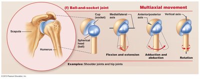
Shoulder Joint
The glenohumeral joint is a ball-and-socket joint, the most freely moving joint in the body. Stability is provided by the articular capsule, coracohumeral and glenohumeral ligaments, the tendon of the long head of the biceps brachii, and the rotator cuff muscles.
Elbow Joint
The elbow is a hinge joint allowing flexion and extension. Stability is provided by the ulna, articular capsule, annular ligament, radial collateral ligament, and ulnar collateral ligament. 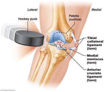
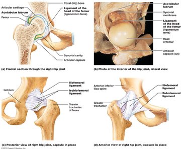
Hip Joint
The hip is a ball-and-socket joint with less range of motion than the shoulder. The femur head fits into the acetabulum, and stability is provided by the articular capsule, iliofemoral, pubofemoral, ischiofemoral ligaments, and ligamentum teres. 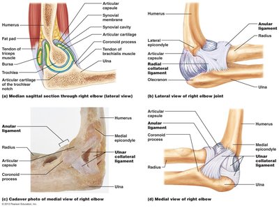
Homeostatic Imbalances of Joints
Common Joint Injuries
Cartilage tears: Often involve the meniscus, caused by compression and shearing stress.
Sprains: Stretching or tearing of ligaments, common in the ankle, knee, and lumbar region.
Dislocations: Bones are forced out of alignment; subluxation is partial dislocation.
Bursitis: Inflammation of the bursa due to trauma or friction.
Tendonitis: Inflammation of the tendon sheath from overuse.
Arthritis and Other Disorders
Osteoarthritis: Degenerative, age-related joint disease.
Rheumatoid arthritis: Chronic inflammatory disorder with long-term effects.
Gouty arthritis: Caused by urate crystal deposits from high uric acid levels, often affecting the big toe.
Lyme Disease: Bacterial infection from tick bites causing joint inflammation.
Summary Tables
Summary of Joint Classes
Structural Class | Structural Characteristics | Types | Mobility |
|---|---|---|---|
Fibrous | Adjoining bones united by collagen fibers | Suture (short fibers), Syndesmosis (longer fibers), Gomphosis (periodontal ligament) | Immobile (suture, gomphosis), Slightly movable (syndesmosis) |
Cartilaginous | Adjoining bones united by cartilage | Synchondrosis (hyaline cartilage), Symphysis (fibrocartilage) | Immobile (synchondrosis), Slightly movable (symphysis) |
Synovial | Adjoining bones covered with articular cartilage, separated by a joint cavity, and enclosed within an articular capsule lined with synovial membrane | Plane, Hinge, Pivot, Condylar, Saddle, Ball-and-socket | Freely movable (diarthrosis; movements depend on design of joints) |
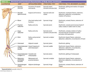
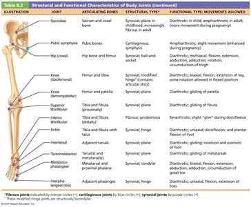
Structural and Functional Characteristics of Body Joints
Joint | Articulating Bones | Structural Type | Functional Type; Movements Allowed |
|---|---|---|---|
Shoulder (glenohumeral) | Scapula and humerus | Synovial; ball-and-socket | Diarthrotic; multiaxial, flexion, extension, abduction, adduction, rotation |
Elbow | Ulna and radius with humerus | Synovial; hinge | Diarthrotic; uniaxial, flexion, extension |
Hip (coxal) | Femur and pelvis | Synovial; ball-and-socket | Diarthrotic; multiaxial, flexion, extension, abduction, adduction, rotation |
Knee (tibiofemoral) | Femur and tibia | Synovial; modified hinge | Diarthrotic; biaxial, flexion, extension, rotation |
Pubic symphysis | Pubic bones | Cartilaginous; symphysis | Amphiarthrotic; slight movement |
Key Equations and Concepts
Joint Movement Axes
Uniaxial: Movement in one plane
Biaxial: Movement in two planes
Multiaxial: Movement in three planes
Example Equation: Range of Motion
Range of motion (ROM) is determined by the structure of the joint and the tension of ligaments and muscles. While there is no single formula, the concept can be summarized as:
Additional info: This equation is a conceptual summary; actual measurement of ROM is done clinically with goniometers.
