 Back
Joints: Structure, Classification, and Function in Human Anatomy & Physiology Ch 9
Study Guide - Smart Notes
 Tailored notes based on your materials, expanded with key definitions, examples, and context.
Joints and Their Classification
Introduction to Joints
Joints, or articulations, are points where two bones meet, regardless of their mobility. The study of joints is called arthrology, while kinesiology focuses on musculoskeletal movement. Joints are named based on the bones involved and are classified by how the bones are bound together: bony, fibrous, cartilaginous, and synovial joints.
Function of joints: Enable movement, provide stability, and allow for growth and development of the skeleton.
Joint classification: Based on the material binding the bones and the presence or absence of a joint cavity.
Bony Joints (Synostosis)
A bony joint is an immobile joint formed when the gap between two bones ossifies, making them a single bone. These can originate from fibrous or cartilaginous joints.
Examples: Fusion of frontal bones in infants, hip bone formation from ilium, ischium, and pubis, and fusion of the first rib to the sternum with age.
Fibrous Joints (Synarthrosis)
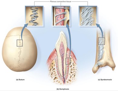
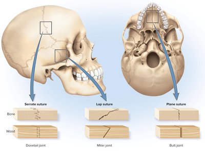
In fibrous joints, adjacent bones are bound by collagen fibers. There are three main types: sutures, gomphoses, and syndesmoses.
Sutures: Immobile or slightly mobile joints found only in the skull, with short collagen fibers binding the bones.
Gomphoses: Attachments of teeth to their sockets, held by the periodontal ligament.
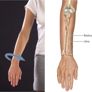
Syndesmoses: Bones are bound by longer collagen fibers, allowing more mobility (e.g., interosseous membrane between radius and ulna).
Cartilaginous Joints (Amphiarthrosis)
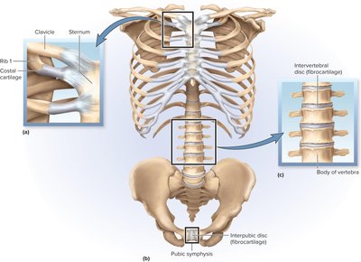
In cartilaginous joints, two bones are linked by cartilage. There are two types: synchondroses and symphyses.
Synchondroses: Bones joined by hyaline cartilage (e.g., epiphyseal plate in children, first rib to sternum).
Symphyses: Bones joined by fibrocartilage (e.g., pubic symphysis, intervertebral discs).
Synovial Joints (Diarthrosis)
General Anatomy of Synovial Joints
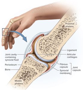
Synovial joints are the most familiar and mobile type of joint, characterized by a joint cavity filled with synovial fluid. They are structurally complex and crucial for movement and quality of life.
Articular cartilage: Hyaline cartilage covering bone surfaces.
Joint (articular) cavity: Space separating the bones, filled with synovial fluid.
Synovial fluid: Lubricates, nourishes cartilage, and removes waste.
Joint capsule: Encloses the cavity, with an outer fibrous layer and inner synovial membrane.
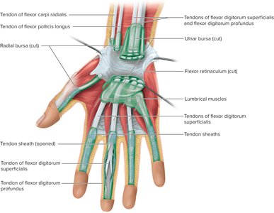
Accessory structures: Tendons (muscle to bone), ligaments (bone to bone), bursae (fluid-filled sacs), and tendon sheaths (elongated bursae around tendons).
Exercise and Articular Cartilage
Exercise increases synovial fluid absorption by cartilage, improving cushioning and nutrient delivery. Lack of exercise leads to cartilage deterioration due to poor nutrition and waste removal.
Joints and Lever Systems
Levers in the Body
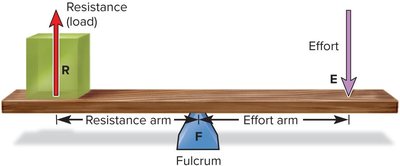
Long bones act as levers to enhance movement. A lever is a rigid object that rotates around a fulcrum. In the body, joints are fulcrums, muscles provide effort, and the object moved is the resistance.
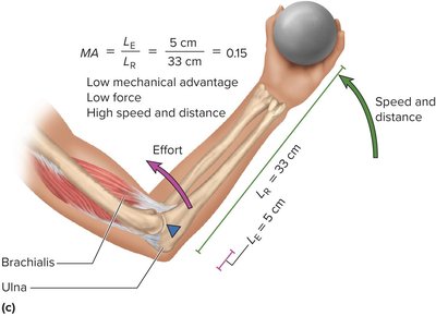
Effort arm: Distance from fulcrum to point of effort.
Resistance arm: Distance from fulcrum to point of resistance.
Mechanical Advantage (MA)
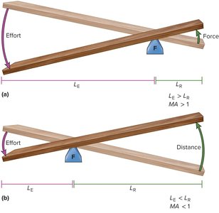
Mechanical advantage is the ratio of output force to input force, calculated as:
If MA > 1: More force, less speed/distance.
If MA < 1: More speed/distance, less force.
Classes of Levers
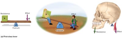
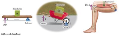
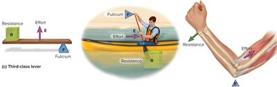
There are three classes of levers, classified by the arrangement of fulcrum (F), effort (E), and resistance (R):
First-class (EFR): Fulcrum in the middle (e.g., atlanto-occipital joint).
Second-class (FRE): Resistance in the middle (e.g., standing on tiptoe).
Third-class (REF): Effort in the middle (e.g., biceps curl).
Range of Motion and Axes of Rotation
Range of Motion (ROM)
Range of motion is the degree through which a joint can move. It is determined by the structure of articular surfaces, ligament and capsule tautness, and muscle/tendon action.
Muscle tone: State of tension in resting muscles, monitored by the nervous system.
Axes of Rotation
Joints can be classified by the number of axes of rotation:
Multiaxial: Three axes (e.g., shoulder joint).
Biaxial: Two axes.
Monoaxial: One axis.
Classes of Synovial Joints
Types of Synovial Joints
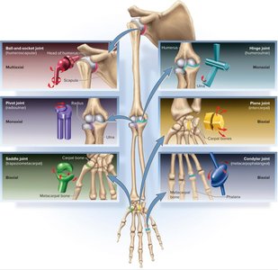
There are six major types of synovial joints, classified by shape and degrees of freedom:
Ball-and-socket: Multiaxial (shoulder, hip).
Condylar (ellipsoid): Biaxial (radiocarpal, metacarpophalangeal).
Saddle: Biaxial (thumb, sternoclavicular).
Plane (gliding): Biaxial (carpals, tarsals, vertebral processes).
Hinge: Monoaxial (elbow, knee, fingers, toes).
Pivot: Monoaxial (atlantoaxial, radioulnar).
Movements of Synovial Joints
Basic Movements
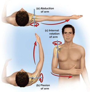
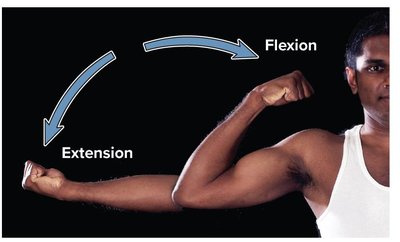
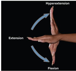
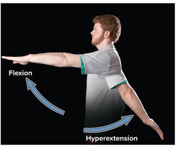
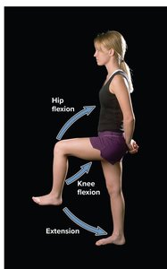
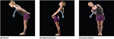
Flexion: Decreases joint angle.
Extension: Increases joint angle.
Hyperextension: Extension beyond anatomical position.
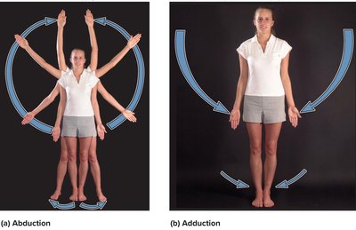
Abduction: Movement away from midline.
Adduction: Movement toward midline.
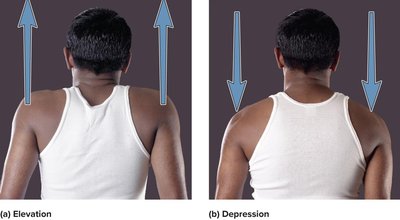
Elevation/Depression: Raising/lowering a body part.
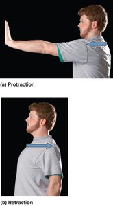
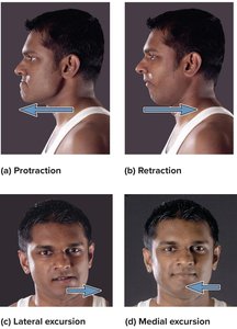
Protraction/Retraction: Anterior/posterior movement in the transverse plane.
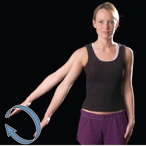
Circumduction: Circular movement of a limb.
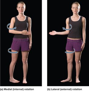
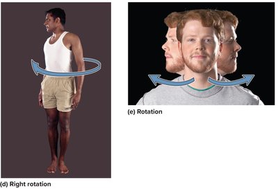
Rotation: Bone spins on its axis (medial/lateral).
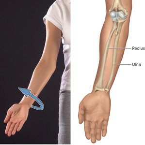
Supination/Pronation: Rotational movement of the forearm or foot.
Anatomy of Selected Diarthroses
Jaw Joint (Temporomandibular Joint, TMJ)
The TMJ is a synovial joint between the condyle of the mandible and the mandibular fossa of the temporal bone. It contains an articular disc and is supported by ligaments. TMJ dysfunction can cause pain, clicking, and restricted movement.
Shoulder Joint (Glenohumeral Joint)
The shoulder joint is the most mobile joint, with a shallow glenoid cavity and loose capsule. It is stabilized by the rotator cuff muscles, ligaments, and several bursae. Dislocations are common due to its mobility.
Elbow Joint
The elbow is a hinge joint with two articulations (humeroulnar and humeroradial) and a pivot joint (proximal radioulnar). Ligaments restrict side-to-side motion, and the annular ligament holds the radius in place for pronation and supination.
Hip Joint (Coxal Joint)
The hip joint is a deep ball-and-socket joint, stabilized by a fibrocartilage labrum and strong ligaments. Dislocations are rare due to its depth and ligamentous support.
Knee Joint (Tibiofemoral Joint)
The knee is the largest and most complex diarthrosis, primarily a hinge joint but capable of slight rotation and gliding. It contains menisci, collateral and cruciate ligaments, and numerous bursae. Injuries often involve the menisci or ACL.
Ankle Joint (Talocrural Joint)
The ankle includes two articulations, stabilized by strong ligaments and the Achilles tendon. Sprains are common due to the range of motion and ligamentous structure.
Arthritis and Artificial Joints
Types of Arthritis
Osteoarthritis (OA): Degenerative "wear-and-tear" arthritis, leading to cartilage breakdown and bone spurs.
Rheumatoid Arthritis (RA): Autoimmune disorder attacking joint tissues, leading to inflammation, cartilage destruction, and joint fusion (ankylosis).
Arthroplasty is the surgical replacement of a diseased joint with an artificial prosthesis.
Joint Type | Binding Material | Mobility | Examples |
|---|---|---|---|
Bony (Synostosis) | Bone | Immobile | Frontal bone fusion |
Fibrous (Synarthrosis) | Collagen fibers | Immobile/slightly mobile | Sutures, gomphoses, syndesmoses |
Cartilaginous (Amphiarthrosis) | Cartilage | Slightly mobile | Epiphyseal plate, pubic symphysis |
Synovial (Diarthrosis) | Synovial cavity | Freely mobile | Shoulder, knee, hip |
Additional info: This summary includes expanded academic context and examples for clarity and completeness, as well as a comparative table of joint types.
