 Back
BackJoints: Structure, Classification, and Movements
Study Guide - Smart Notes
 Tailored notes based on your materials, expanded with key definitions, examples, and context.
Joints
Functional Classification of Joints
Joints, also known as articulations, are classified functionally based on the amount of movement they allow. This classification helps in understanding their role in the human body.
Synarthroses: Immovable joints, typically found in areas requiring stability.
Amphiarthroses: Slightly movable joints, providing a balance between stability and mobility.
Diarthroses: Freely movable joints, essential for most body movements.
Structural Classification of Joints
Joints are also classified structurally based on the material binding the bones and the presence or absence of a joint cavity.
Fibrous Joints: Bones are joined by fibrous tissue; most are immovable.
Cartilaginous Joints: Bones are united by cartilage. These joints allow more movement than fibrous joints but less than synovial joints.
Synovial Joints: Bones are separated by a fluid-filled joint cavity, allowing free movement.
Cartilaginous Joints
Cartilaginous joints are further divided into two types: synchondroses and symphyses.
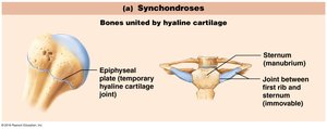
Synchondroses
Synchondroses are joints where bones are united by hyaline cartilage. These joints are typically immovable.
Epiphyseal plate: Temporary joint found in growing bones.
Sternum (manubrium): Joint between the first rib and sternum is immovable.
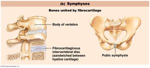
Symphyses
Symphyses are joints where bones are united by fibrocartilage, allowing limited movement.
Intervertebral discs: Fibrocartilaginous discs between vertebrae provide cushioning and flexibility.
Pubic symphysis: Joint between the pubic bones in the pelvis.
Synovial Joints
Synovial joints are the most common and freely movable joints in the body. They have a complex structure that allows a wide range of movements.
Articular cartilage: Covers the ends of bones, reducing friction and absorbing shock.
Joint cavity: Space containing synovial fluid.
Synovial fluid: Lubricates the joint, nourishes cartilage, and reduces friction.
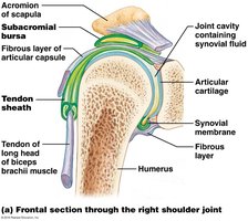
Synovial Joint Structure
Articular capsule: Encloses the joint cavity; consists of a fibrous layer and a synovial membrane.
Fibrous layer: Provides strength and stability.
Synovial membrane: Produces synovial fluid.
Reinforcing ligaments: Strengthen and support the joint.
Nerves and blood vessels: Supply the joint, aiding in function and repair.
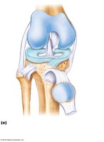
Articular Discs (Menisci)
Articular discs, or menisci, are pads of fibrocartilage that improve the fit between articulating bone surfaces, stabilize joints, and minimize wear and tear.
Stabilizes joints: Distributes load and reduces stress.
Minimizes wear and tear: Protects articular cartilage.
Bursae
Bursae are fluid-filled sacs lined with synovial membrane, found in areas of friction such as between tendons and bones. They help reduce friction and facilitate movement.
Structure: Lined with synovial membrane and contain a thin film of synovial fluid.
Body Movements at Synovial Joints
Synovial joints allow a variety of movements, each with specific terminology:
Flexion & Extension: Decreasing and increasing the angle between bones, respectively.
Abduction & Adduction: Moving a limb away from or toward the body's midline.
Circumduction & Rotation: Moving a limb in a circular motion or rotating it around its axis.
Supination & Pronation: Rotating the forearm to turn the palm up or down.
Inversion & Eversion: Turning the sole of the foot inward or outward.
Dorsiflexion & Plantar Flexion: Moving the foot upward or downward at the ankle.
Protraction & Retraction: Moving a body part forward or backward.
Elevation & Depression: Raising or lowering a body part.
Example: The shoulder joint, a synovial joint, allows flexion, extension, abduction, adduction, circumduction, and rotation.
Additional info: Synovial joints are essential for most voluntary movements and are commonly affected by injuries and diseases such as arthritis.
