 Back
Ch 9: Joints: Structure, Function, and Movement
Study Guide - Smart Notes
 Tailored notes based on your materials, expanded with key definitions, examples, and context.
Joints: Structure, Function, and Movement
Introduction to Joints
Joints, or articulations, are connections between bones that allow for varying degrees of movement and stability. They are classified by both their anatomical structure and their functional range of motion. Understanding the relationship between joint structure and function is essential for comprehending how the skeletal system supports movement and stability.
Classification of Joints
Structural and Functional Classification
Joints are categorized by their structure (the type of tissue connecting the bones) and by their function (the degree of movement they allow):
Structural Classification: Fibrous, cartilaginous, bony, and synovial joints.
Functional Classification: Synarthrosis (immovable), amphiarthrosis (slightly movable), and diarthrosis (freely movable).
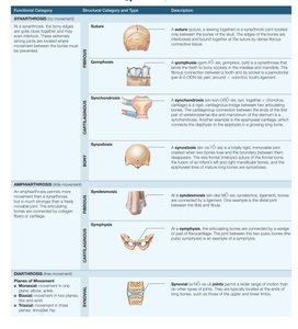
These classifications are summarized in the following table:
Functional Category | Structure Category and Type | Description |
|---|---|---|
Synarthrosis (Immovable) | Suture, Gomphosis, Synchondrosis, Synostosis | Sutures are found between skull bones; gomphoses anchor teeth; synchondroses are cartilaginous (e.g., epiphyseal plates); synostoses are fused bones (e.g., frontal bone). |
Amphiarthrosis (Slightly Movable) | Syndesmosis, Symphysis | Syndesmoses are connected by ligaments (e.g., distal tibiofibular joint); symphyses are connected by fibrocartilage (e.g., pubic symphysis). |
Diarthrosis (Freely Movable) | Synovial | Permit a wide range of motion; all synovial joints are diarthroses (e.g., shoulder, knee). |
Synovial Joints (Diarthroses)
Basic Structure of Synovial Joints
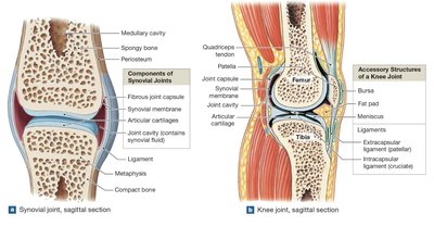
Synovial joints are characterized by a joint cavity filled with synovial fluid and surrounded by a joint capsule. These joints are highly mobile but less stable than other types. The main components include:
Articular Cartilage: Covers the articulating surfaces, reducing friction and absorbing shock.
Joint Capsule: A two-layered structure with an outer fibrous capsule and an inner synovial membrane.
Synovial Fluid: Lubricates, nourishes, and cushions the joint.
Accessory Structures of Synovial Joints
Menisci: Pads of fibrocartilage that improve fit and channel synovial fluid.
Fat Pads: Masses of adipose tissue that protect and fill spaces as the joint moves.
Ligaments: Strengthen and reinforce the joint; can be extracapsular or intracapsular.
Tendons: Connect muscle to bone, stabilizing the joint.
Bursae: Fluid-filled sacs that reduce friction and absorb shock where tendons or ligaments rub against other tissues.
Stability and Mobility
There is an inverse relationship between joint mobility and stability. Highly mobile joints (e.g., shoulder) are less stable, while less mobile joints (e.g., hip) are more stable due to structural reinforcements.
Movements at Synovial Joints
Types of Movements
Movements at synovial joints are described using precise anatomical terms:
Gliding: Sliding of flat surfaces (e.g., between carpal bones).
Angular Movements:
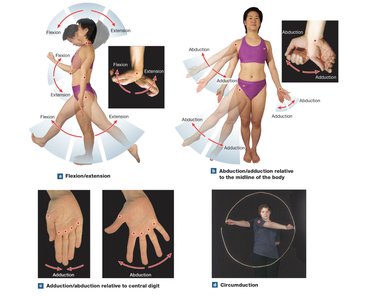
Flexion: Decreases the angle between bones.
Extension: Increases the angle between bones.
Hyperextension: Extension beyond the anatomical position.
Abduction: Movement away from the midline.
Adduction: Movement toward the midline.
Circumduction: Circular movement combining flexion, extension, abduction, and adduction.
Rotational Movements:
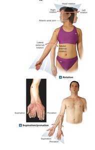
Medial (Internal) Rotation: Anterior surface turns toward the midline.
Lateral (External) Rotation: Anterior surface turns away from the midline.
Pronation: Palm turns posteriorly.
Supination: Palm turns anteriorly (anatomical position).
Special Movements:
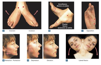
Inversion/Eversion: Twisting the foot inward/outward.
Dorsiflexion/Plantar Flexion: Flexing/extending the ankle.
Opposition/Reposition: Moving the thumb toward/away from the fingers.
Protraction/Retraction: Moving a body part anteriorly/posteriorly.
Elevation/Depression: Moving a structure superiorly/inferiorly.
Lateral Flexion: Bending the vertebral column to the side.
Classification of Synovial Joints by Movement
Synovial joints are further classified by the axes and types of movement they permit:
Plane (Gliding) Joints: Permit sliding movements (e.g., intercarpal joints).
Hinge Joints: Permit flexion and extension (e.g., elbow).
Condylar (Ellipsoid) Joints: Permit angular movement in two planes (e.g., wrist).
Saddle Joints: Permit angular movement, including opposition (e.g., thumb).
Pivot Joints: Permit rotation (e.g., proximal radioulnar joint).
Ball-and-Socket Joints: Permit the greatest range of motion (e.g., shoulder, hip).
Degenerative Changes and Aging
Effects of Aging on Joints
With age, joints are prone to degenerative changes, leading to reduced mobility and increased risk of injury. Common age-related joint problems include:
Osteoarthritis: Degenerative joint disease due to wear and tear or genetic factors.
Rheumatoid Arthritis: Autoimmune inflammation of joint tissues.
Gouty Arthritis: Deposition of uric acid crystals in synovial fluid, often affecting the great toe.
Other changes include decreased bone mass, increased risk of fractures, and possible joint immobilization leading to cartilage degeneration. Regular exercise, physical therapy, and anti-inflammatory drugs can help manage symptoms and slow progression.
Summary Table: Types of Joints
Type | Structure | Movement | Example |
|---|---|---|---|
Synarthrosis | Fibrous or cartilaginous | Immovable | Sutures of skull |
Amphiarthrosis | Fibrous or cartilaginous | Slightly movable | Pubic symphysis |
Diarthrosis (Synovial) | Synovial cavity, capsule, fluid | Freely movable | Shoulder, knee |
Additional info: The above content integrates and expands upon the provided materials, ensuring a comprehensive, self-contained study guide for exam preparation in anatomy and physiology.
