 Back
BackKnee Joint Anatomy Labeling Guidance
Study Guide - Smart Notes
 Tailored notes based on your materials, expanded with key definitions, examples, and context.
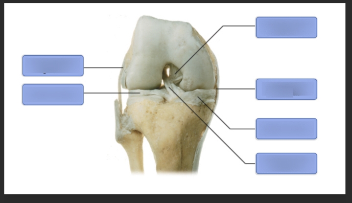
Q1. Label the anatomical structures of the knee joint shown in the image.
Background
Topic: Musculoskeletal Anatomy – Knee Joint
This question tests your knowledge of the anatomical features of the knee joint, including bones, ligaments, and cartilage. Understanding these structures is essential for ANP (Anatomy and Physiology) college courses, especially for topics related to joint function, injury, and movement.
Key Terms:
Femur: The thigh bone, forming the upper part of the knee joint.
Tibia: The shin bone, forming the lower part of the knee joint.
Patella: The kneecap, located anteriorly.
Meniscus: Cartilaginous structures that cushion and stabilize the joint.
Ligaments: Connective tissues that stabilize the knee (e.g., ACL, PCL, MCL, LCL).
Step-by-Step Guidance
Observe the image and note the orientation: The femur is at the top, tibia at the bottom, and the patella is not visible in this posterior view.
Identify the major bones: The upper bone is the femur, and the lower bone is the tibia.
Locate the menisci: These are the crescent-shaped cartilages between the femur and tibia, visible as darkened areas.
Identify the ligaments: The central structures crossing between the femur and tibia are likely the anterior and posterior cruciate ligaments (ACL and PCL).
Match each label box to the corresponding anatomical structure based on its position in the image.
Try solving on your own before revealing the answer!
