 Back
BackLab 1: Organization, Tissues, and the Integument – ANP College Study Notes
Study Guide - Smart Notes
 Tailored notes based on your materials, expanded with key definitions, examples, and context.
Overview of Anatomy and Physiology
Introduction to Anatomy and Physiology
Anatomy is the study of the structure of the human body, often revealed through dissection, while physiology focuses on the functions of these structures and their role in maintaining homeostasis. Mastery of anatomy and physiology requires learning precise terminology, much of which is derived from Latin and Greek roots.
Anatomy: Examines body structures and their relationships.
Physiology: Explores how anatomical structures function together.
Homeostasis: The body's ability to maintain a stable internal environment.
Importance of Terminology: Accurate communication in anatomy relies on standardized terms.
Anatomical Terminology
Anatomical Position and Body Regions
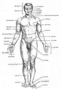
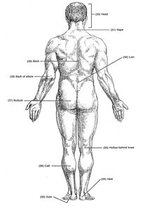
The anatomical position is the standard reference for describing body regions. The subject stands erect, facing forward, with arms at the sides and palms facing forward. Anatomical terms are used to describe regions and structures precisely.
Anatomical Position: Standing, facing observer, arms at sides, palms forward, feet flat.
Regional Terminology: Terms like brachial (arm), olecranal (elbow), and femoral (thigh) describe specific areas.
Adjectives: Used to modify structure names based on location (e.g., brachial artery).
Example: The olecranal region refers to the elbow area, specifically the superior portion of the ulna.
Directional Terms
Directional terms provide clarity when describing the location of body structures relative to each other.
Superior: Toward the head or upper part.
Inferior: Toward the feet or lower part.
Anterior: Toward the front.
Posterior: Toward the back.
Medial: Toward the midline.
Lateral: Away from the midline.
Intermediate: Between two structures.
Ipsilateral: Same side.
Contralateral: Opposite side.
Proximal: Nearer to the point of attachment.
Distal: Farther from the point of attachment.
Superficial: Close to the surface.
Deep: Further from the surface.
Planes of the Body
Body planes are imaginary flat surfaces used to divide the body and describe anatomical relationships.
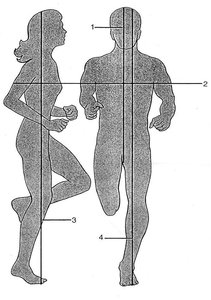
Midsagittal (Median) Plane: Divides the body into equal right and left halves.
Parasagittal Plane: Divides the body into unequal right and left sides.
Frontal (Coronal) Plane: Divides the body into anterior and posterior portions.
Transverse (Horizontal) Plane: Divides the body into superior and inferior portions.
Body Cavities
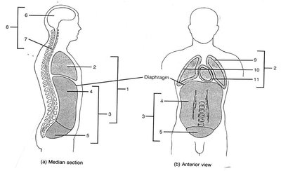
Body cavities are spaces within the body that house and protect internal organs. They are classified as posterior or anterior cavities, each containing specific organs.
Posterior Body Cavity: Includes the cranial cavity (brain) and vertebral cavity (spinal cord).
Anterior Body Cavity: Divided by the diaphragm into thoracic and abdominopelvic cavities.
Thoracic Cavity: Contains pleural cavities (lungs), mediastinum (heart, thymus, esophagus, trachea), and pericardial cavity (heart).
Abdominopelvic Cavity: Includes abdominal (digestive organs) and pelvic (bladder, reproductive organs) cavities.
Quadrants: Abdominopelvic cavity is divided into four quadrants for clinical reference.
Levels of Biological Organization
Hierarchy of Organization
The human body is organized into hierarchical levels, each contributing to overall function and homeostasis.
Chemical Level: Atoms and molecules form the basis of life.
Cellular Level: Cells are the basic units of structure and function.
Tissue Level: Groups of similar cells perform specific functions.
Organ Level: Organs consist of multiple tissue types working together.
System Level: Organ systems coordinate to maintain homeostasis.
Organism Level: The complete living individual.
Example: The respiratory system exchanges gases, supporting the cardiovascular system in oxygen transport and carbon dioxide removal.
Additional info: Disruption of homeostasis can result from disease, trauma, or environmental changes.
Primary Tissue Types
Classification of Tissues
Tissues are classified into four main types: epithelial, connective, nervous, and muscular. Each type has unique structural and functional characteristics.
Epithelial Tissue: Covers surfaces, lines cavities, forms glands.
Connective Tissue: Supports, binds, protects, and transports substances.
Muscular Tissue: Produces movement through contraction.
Nervous Tissue: Transmits electrical signals for communication.
Key Terms: cyte (mature cell), blast (immature cell), fibroblast (fiber-producing cell), chondroblast (cartilage-producing cell).
Epithelial Tissues
Epithelial tissues perform protection, absorption, secretion, filtration, and sensory functions. They are classified by cell shape and number of layers.
Simple Epithelium: One cell layer; allows rapid diffusion.
Stratified Epithelium: Multiple layers; provides protection.
Cell Shapes: Squamous (flat), cuboidal (cube-shaped), columnar (tall).
Pseudostratified Columnar Epithelium: Appears layered but is a single layer; often ciliated.
Epithelium Type | Function | Location |
|---|---|---|
Simple Squamous | Diffusion, filtration | Alveoli, blood vessels, body cavities |
Simple Cuboidal | Secretion, absorption | Kidney tubules |
Simple Columnar | Absorption, secretion | Small intestine |
Pseudostratified Columnar | Secretion, movement of mucus | Trachea, respiratory tract |
Stratified Squamous | Protection against abrasion | Epidermis, mouth, esophagus, anus, vagina |
Connective Tissue
Connective tissues provide structural support, bind tissues, protect organs, and transport substances. They originate from mesenchyme and vary in ground substance and fiber composition.
Hyaline Cartilage: Firm, flexible; found at ends of long bones, nose, trachea.
Loose Connective Tissue (Areolar): Packing material; beneath skin, around organs.
Dense Regular Connective Tissue: Parallel collagen fibers; tendons, ligaments.
Dense Irregular Connective Tissue: Random collagen fibers; dermis of skin.
Elastic Connective Tissue: Elastic fibers; large blood vessels.
Adipose Tissue: Fat storage; beneath skin, around organs.
Connective Tissue Type | Function | Location |
|---|---|---|
Hyaline Cartilage | Support, flexibility | Ends of long bones, nose, trachea |
Loose Connective | Binding, support | Beneath skin, around organs |
Dense Regular | Strength in one direction | Tendons, ligaments |
Dense Irregular | Strength in multiple directions | Dermis of skin |
Elastic Connective | Expansion, recoil | Large blood vessels |
Adipose | Energy storage, insulation, protection | Beneath skin, around organs |
Example: Tendons and ligaments are made of dense regular connective tissue for strength and resistance to tension in one direction.
Additional info: Adipose tissue functions as energy storage, thermal insulation, and organ protection.
Nervous Tissue
Nervous tissue consists of neurons and neuroglia. Neurons transmit electrical signals, while neuroglia support and protect neurons.
Neurons: Excitable cells with axons (transmit signals) and dendrites (receive signals).
Neuroglia: Supportive cells; maintain environment, anchor neurons, facilitate impulse transmission.
Nervous Tissue Type | Function | Location |
|---|---|---|
Multipolar Neuron | Signal transmission | Brain, spinal cord, peripheral nerves |
Muscular Tissue
Muscle tissue is specialized for contraction and movement. It is classified into skeletal, cardiac, and smooth muscle.
Skeletal Muscle: Voluntary, striated, multinucleated; attached to skeleton.
Cardiac Muscle: Involuntary, striated, branched; found in heart, single nucleus, intercalated discs.
Smooth Muscle: Involuntary, non-striated; found in organs, blood vessels, iris.
Muscle Tissue Type | Function | Location |
|---|---|---|
Skeletal Muscle | Movement | Attached to bones |
Cardiac Muscle | Pumping blood | Heart |
Smooth Muscle | Movement in organs | Digestive tract, blood vessels, iris |
The Integumentary System
Structure and Function of the Integument
The integumentary system protects the body, regulates temperature, excretes waste, and provides sensation. It consists of the epidermis, dermis, and hypodermis.
Epidermis: Stratified squamous epithelium; contains keratinocytes producing keratin for resilience.
Dermis: Dense irregular connective tissue; contains hair follicles, glands, sensory receptors.
Hypodermis: Adipose and connective tissue; provides insulation and energy storage.
Glands: Sebaceous (oil) and eccrine (sweat) glands; sebum prevents desiccation, sweat regulates temperature and provides antimicrobial protection.
Hair: Keratinized cells; protection, sensation.
Arrector Pili Muscle: Moves hair upright in response to cold or fear.
Pacinian Corpuscle: Sensory receptor for pressure and touch.
Layers of the Epidermis:
Stratum Corneum: Outermost, keratinized cells.
Stratum Lucidum: Present in thick skin (soles, palms).
Stratum Granulosum: Produces keratin and lipids for water barrier.
Stratum Spinosum: Provides strength and flexibility.
Stratum Basale: Deepest, mitotically active stem cells.
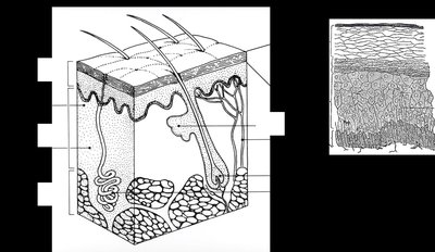
Example: The stratum corneum protects against abrasion, while the lipid layer in the stratum granulosum prevents water loss.
Additional info: The epidermis relies on diffusion from the dermis for nutrients and oxygen due to its avascularity.
