 Back
BackLab 5 Study Guide: Articulations, Muscle Anatomy, and Muscle Functions
Study Guide - Smart Notes
 Tailored notes based on your materials, expanded with key definitions, examples, and context.
Articulations (Joints) Anatomy
Knee, Hip, and Shoulder Joints
Articulations, or joints, are anatomical locations where two bones meet, often allowing movement. The knee, hip, and shoulder are major synovial joints with complex structures that provide stability and mobility.
Knee Joint: Includes the Anterior Cruciate Ligament, Posterior Cruciate Ligament, Medial Meniscus, Lateral Meniscus, Patellar Ligament, Fibular (Lateral) Collateral Ligament, and Tibial (Medial) Collateral Ligament.
Hip Joint: Features the Pubofemoral Ligament, Iliofemoral Ligament, and Ischiofemoral Ligament.
Shoulder Joint: Contains the Coracoacromial Ligament, Glenohumeral Ligament, Coracoclavicular Ligament, and Acromioclavicular Ligament.
Example: The knee joint is stabilized by both cruciate and collateral ligaments, which prevent excessive movement and injury.
Microscopic Anatomy of Muscle Cells
Sarcomere Structure and Muscle Cell Components
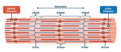
Skeletal muscle contraction relies on the organization of microscopic structures within muscle cells. The sarcomere is the fundamental contractile unit, composed of repeating bands and lines that organize actin and myosin filaments.
Sarcomere: The basic unit of muscle contraction, defined by Z discs.
Z Disc: Boundary of the sarcomere; anchors actin filaments.
M Line: Center of the sarcomere; anchors myosin filaments.
A Band: Region containing the entire length of myosin filaments.
I Band: Region containing only actin filaments.
H Zone: Central region of the A band with only myosin filaments.
Sarcolemma: Muscle cell membrane.
Sarcoplasm: Cytoplasm of the muscle cell.
Sarcoplasmic Reticulum: Stores and releases calcium ions for contraction.
Terminal Cisternae: Enlarged areas of the sarcoplasmic reticulum adjacent to T tubules.
Transverse (T) Tubules: Invaginations of the sarcolemma that transmit action potentials.
Mitochondria: Provide ATP for muscle contraction.
Nuclei: Multiple per muscle cell, reflecting its multinucleate nature.
Neuromuscular Junction: Site where motor neuron stimulates muscle cell.
Myofibrils: Bundles of contractile filaments within the muscle cell.
Myofilaments: Actin (thin) and myosin (thick) filaments responsible for contraction.
Actin Myofilaments: Thin filaments anchored to Z discs.
Myosin Myofilaments: Thick filaments anchored to M line.
Example: During contraction, myosin heads bind to actin filaments and pull them toward the M line, shortening the sarcomere.
Skeletal Muscles: Gross Anatomy and Functions
Muscle Groups and Their Functions
Muscles are classified by their location and function. Understanding their actions is essential for identifying their roles in movement and stability.
Muscles of Facial Expression: Occipitofrontalis (frontalis, occipitalis), Buccinator, Orbicularis Oculi, Orbicularis Oris, Mentalis, Platysma, Zygomaticus Major, Zygomaticus Minor.
Muscles of Mastication: Temporalis, Masseter.
Muscles That Move the Head and Neck: Splenius Capitis, Sternocleidomastoid, Scalene.
Muscles of Ventilation: External Intercostals, Internal Intercostals, Diaphragm.
Abdominal Muscles: Rectus Abdominus, Internal Obliques, External Obliques, Transversus Abdominus.
Muscles That Move the Vertebral Column: Erector Spinae Group.
Muscles That Move the Scapula at the Pectoral Girdle: Subclavius, Pectoralis Minor, Levator Scapulae, Serratus Anterior, Trapezius, Rhomboid Major, Rhomboid Minor.
Connective Tissue Elements: Epicranial Aponeurosis, Aponeurosis of External Oblique, Lumbodorsal Fascia, Linea Alba.
Example: The Masseter muscle elevates the mandible, allowing the jaw to close during chewing.
Muscle Functions Table
The following table summarizes the primary functions of key muscles as listed in the lab manual. Only these functions are accepted for exam credit.
Muscle | Function(s) |
|---|---|
Frontalis | Elevates eyebrows; wrinkles skin of forehead |
Occipitalis | Pulls scalp posteriorly |
Buccinator | Compresses the cheek |
Orbicularis Oculi | Closes eye |
Orbicularis Oris | Closes and protrudes the lip |
Mentalis | Protrudes lower lip |
Platysma | Depresses lower lip; tenses skin of neck |
Zygomaticus Major | Pulls angle of mouth superiorly and laterally |
Zygomaticus Minor | Elevates lateral portion of the lip |
Temporalis | Retracts and elevates mandible |
Masseter | Closes jaw; elevates mandible |
Splenius Capitis | Extends, rotates and laterally flexes neck |
Sternocleidomastoid | Flexes neck |
Scalene | Elevates rib cage |
Erector Spinae | Extends vertebral column |
External Intercostals | Elevates rib cage |
Internal Intercostals | Depresses rib cage |
Diaphragm | Expands thoracic cavity; compresses abdominopelvic cavity |
Rectus Abdominus | Compresses abdominal cavity; flexes vertebral column |
Internal Obliques | Flexes or bends spine; compresses abdomen |
External Obliques | Flexes or bends spine; compresses abdomen; depresses ribs |
Transversus Abdominus | Compresses abdominal cavity |
Pectoralis Minor | Elevates ribs; protracts and depresses scapula |
Levator Scapulae | Elevates, rotates and retracts scapula |
Serratus Anterior | Protracts scapula; superior rotation of scapula |
Trapezius | Rotates scapula posteriorly; retracts and depresses scapula |
Subclavius | Protracts and depresses shoulder |
Rhomboid Major | Adducts scapula; performs downward rotation |
Rhomboid Minor | Adducts scapula; performs downward rotation |
Study Tips and Requirements
Lab 5 Expectations
For quiz preparation, students must:
Identify joint components, microscopic muscle cell structures, and muscles/connective tissue elements on lab models.
List all muscle functions exactly as provided in the lab manual tables.
Use only lab manual definitions and functions; outside sources are not accepted.
Spelling and complete names are required for full credit.
Additional info: Understanding the anatomical terminology and muscle functions is essential for success in anatomy and physiology courses, as these concepts form the basis for clinical and physiological applications.
