 Back
BackLab Manual Study Guide: Brain Anatomy & Cranial Nerves
Study Guide - Smart Notes
 Tailored notes based on your materials, expanded with key definitions, examples, and context.
Q1. What is the superficial layer of the cerebrum called?
Background
Topic: Brain Anatomy
This question tests your knowledge of the structural organization of the cerebrum, specifically the outermost layer that is involved in higher-order brain functions.
Key Terms:
Cerebrum: The largest part of the brain, responsible for voluntary activities, sensation, thought, and memory.
Superficial layer: The outermost layer of a structure.
Step-by-Step Guidance
Recall that the cerebrum is covered by a thin, highly folded layer of gray matter.
This layer is responsible for processing sensory information, reasoning, and voluntary muscle activity.
Think about the term used to describe the outermost layer of the cerebrum, which is also involved in conscious thought.
Try solving on your own before revealing the answer!
Q2. Visual areas are located in which cerebral lobe?
Background
Topic: Functional Brain Regions
This question is about the localization of sensory processing in the brain, specifically where visual information is interpreted.
Key Terms:
Cerebral lobe: One of the major subdivisions of the cerebrum, each with specialized functions.
Visual areas: Regions of the brain responsible for processing visual information from the eyes.
Step-by-Step Guidance
Recall the four main lobes of the cerebrum: frontal, parietal, temporal, and occipital.
Think about which lobe is located at the back of the brain and is primarily responsible for vision.
Identify the lobe that contains the primary visual cortex.
Try solving on your own before revealing the answer!
Q3. Name the fissure that separates the right and left cerebral hemispheres.
Background
Topic: Brain Anatomy – Major Landmarks
This question focuses on the anatomical features that divide the brain into two hemispheres.
Key Terms:
Fissure: A deep groove in the brain's surface.
Cerebral hemispheres: The two halves of the cerebrum (right and left).
Step-by-Step Guidance
Recall the name of the deep groove that runs along the midline of the brain, separating the two hemispheres.
This fissure is visible from a medial view of the brain.
Think about the term that means "longitudinal" or "along the length" of the brain.
Try solving on your own before revealing the answer!
Q4. The optic chiasma gives rise to the optic nerves. Which cranial nerve lies directly anterior to the optic chiasma?
Background
Topic: Cranial Nerves and Brain Anatomy
This question tests your understanding of the spatial relationships of cranial nerves at the base of the brain.
Key Terms:
Optic chiasma: The X-shaped structure where the optic nerves cross.
Cranial nerves: Twelve pairs of nerves that emerge directly from the brain.
Step-by-Step Guidance
Recall the numbering and names of the cranial nerves, especially those near the optic chiasma.
Think about which cranial nerve is located just in front (anterior) of the optic chiasma.
Consider the function of this nerve—it is responsible for the sense of smell.
Try solving on your own before revealing the answer!
Q5. Name the three meningeal layers from most superficial to deepest.
Background
Topic: Protective Coverings of the Brain
This question is about the three connective tissue membranes that surround the brain and spinal cord.
Key Terms:
Meninges: The three layers of protective tissue covering the brain and spinal cord.
Superficial: Closest to the outside.
Deepest: Closest to the brain tissue.
Step-by-Step Guidance
Recall the names of the three meningeal layers, starting from the outermost to the innermost.
Think about the order: dura mater, arachnoid mater, and pia mater.
List them in order from superficial to deep.
Try solving on your own before revealing the answer!
Q6. State two functions of cerebrospinal fluid.
Background
Topic: Brain Physiology
This question tests your understanding of the roles of cerebrospinal fluid (CSF) in the central nervous system.
Key Terms:
Cerebrospinal fluid (CSF): A clear fluid found in the brain and spinal cord.
Functions: The roles or purposes of a structure or substance.
Step-by-Step Guidance
Think about how CSF protects the brain physically and chemically.
Consider its role in cushioning the brain and removing waste products.
List two distinct functions of CSF.
Try solving on your own before revealing the answer!
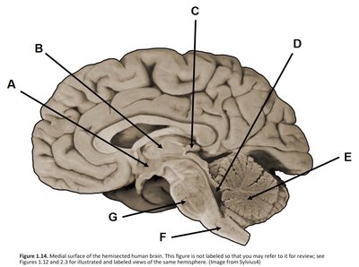
Q7. Study this image of a human brain and identify the labeled parts (A–G).
Background
Topic: Brain Anatomy – Medial View
This question asks you to identify major structures of the brain from a medial (side) view. Use your knowledge of brain anatomy and the typical locations of key structures.
Key Terms:
Corpus callosum, thalamus, hypothalamus, cerebellum, brainstem, etc.
Step-by-Step Guidance
For each arrow (A–G), observe its location and the structure it points to.
Recall the typical position of major brain structures in a sagittal (side) view.
Match each arrow to the most likely anatomical structure based on its position (e.g., A may point to the corpus callosum, B to the thalamus, etc.).
Use your textbook or lab manual diagrams for reference if needed.
Try solving on your own before revealing the answer!
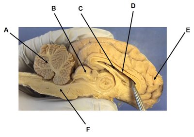
Q8. Study this image of a sheep brain and identify the labeled parts (A–F).
Background
Topic: Comparative Brain Anatomy
This question asks you to identify structures in a dissected sheep brain, which is commonly used in anatomy labs due to its similarity to the human brain.
Key Terms:
Cerebellum, corpus callosum, ventricles, lobes, brainstem, etc.
Step-by-Step Guidance
For each arrow (A–F), observe the structure it points to and compare with diagrams of the sheep brain.
Identify distinguishing features such as the tree-like appearance of the cerebellum (arbor vitae), the curved shape of the corpus callosum, and the location of the ventricles.
Use your lab manual or textbook for labeled images to help match each arrow to the correct structure.
Try solving on your own before revealing the answer!
Unit 16: Cranial Nerves – Clinical Scenarios
Background
Topic: Cranial Nerve Function and Testing
These questions test your ability to associate clinical tests with the correct cranial nerve(s) based on their function.
Key Terms:
Cranial nerves: Twelve pairs of nerves that emerge from the brain and control sensory and motor functions of the head and neck.
Clinical testing: Procedures used to assess the function of specific nerves.
Step-by-Step Guidance
For each scenario, identify the action being tested (e.g., tongue movement, vision, smell, eye movement, facial expression, hearing).
Recall which cranial nerve(s) are responsible for that function (e.g., hypoglossal for tongue, optic for vision, olfactory for smell, oculomotor/trochlear/abducens for eye movement, facial for expression, vestibulocochlear for hearing).
Match the scenario to the correct cranial nerve by name and number.
Try solving on your own before revealing the answer!
Final Answers
Q1: Cerebral cortex
Q2: Occipital lobe
Q3: Longitudinal fissure
Q4: Olfactory nerve (Cranial Nerve I)
Q5: Dura mater, arachnoid mater, pia mater
Q6: 1) Cushions/protects the brain; 2) Removes waste products
Q7: A–G: (A) Corpus callosum, (B) Thalamus, (C) Pineal gland, (D) Midbrain, (E) Pons, (F) Medulla oblongata, (G) Cerebellum
Q8: A–F: (A) Arbor vitae, (B) Corpus callosum, (C) Lateral ventricle, (D) Thalamus, (E) Occipital lobe, (F) Brainstem
Unit 16: 1) Hypoglossal nerve (XII), 2) Optic nerve (II), 3) Olfactory nerve (I), 4) Oculomotor (III), trochlear (IV), abducens (VI), 5) Facial nerve (VII), 6) Vestibulocochlear nerve (VIII)
These answers are based on standard anatomical and physiological knowledge. Review your lab manual for diagrams and further explanations!
