 Back
BackLab Session 8: Eye and Ear (Unit 17) – Study Guidance
Study Guide - Smart Notes
 Tailored notes based on your materials, expanded with key definitions, examples, and context.
Q1. Which three cranial nerves innervate the extrinsic eye muscles?
Background
Topic: Cranial Nerves and Eye Movement
This question tests your knowledge of the cranial nerves responsible for controlling the movement of the extrinsic muscles of the eye, which allow the eyeball to move in different directions.
Key Terms:
Extrinsic eye muscles: Muscles attached to the outside of the eyeball that control its movement.
Cranial nerves: Nerves that emerge directly from the brain and brainstem.
Step-by-Step Guidance
Recall that there are six extrinsic eye muscles responsible for moving the eyeball.
Think about which cranial nerves are involved in controlling these muscles. There are three specific cranial nerves that innervate these muscles.
Review the names and numbers of these cranial nerves in your lab manual or textbook.
Try solving on your own before revealing the answer!
Q2. Where, specifically, is the blind spot located?
Background
Topic: Anatomy of the Eye
This question is about the location of the blind spot, which is an area on the retina where no photoreceptors are present.
Key Terms:
Blind spot: The area of the retina where the optic nerve exits the eye, lacking photoreceptors.
Optic disk: The anatomical term for the blind spot.
Step-by-Step Guidance
Recall that the blind spot is where the optic nerve leaves the eye.
Think about the specific location on the retina where this occurs.
Review the diagram of the eye to identify the optic disk.
Try solving on your own before revealing the answer!
Q3. What does it mean to have “20/70” vision?
Background
Topic: Visual Acuity
This question tests your understanding of how vision is measured and what the numbers in a visual acuity score represent.
Key Terms:
Visual acuity: The clarity or sharpness of vision.
Snellen chart: The chart used to measure visual acuity.
Step-by-Step Guidance
Recall that the first number (20) refers to the distance in feet at which the test is performed.
The second number (70) refers to the distance at which a person with normal vision can read the same line.
Think about what this means for the clarity of vision compared to normal.
Try solving on your own before revealing the answer!
Q4. Define astigmatism.
Background
Topic: Eye Disorders
This question is about the definition and cause of astigmatism, a common vision problem.
Key Terms:
Astigmatism: A condition where the cornea or lens is irregularly shaped, causing blurred vision.
Cornea: The transparent front part of the eye.
Step-by-Step Guidance
Recall that astigmatism is related to the shape of the cornea or lens.
Think about how this irregular shape affects the way light is focused on the retina.
Review the definition in your lab manual for a concise explanation.
Try solving on your own before revealing the answer!
Q6. In mm, record the near point of accommodation for your:
Background
Topic: Accommodation and Vision Testing
This question asks you to measure the closest point at which your eye can focus on an object, known as the near point of accommodation.
Key Terms:
Near point of accommodation: The minimum distance at which the eye can focus on an object.
Accommodation: The process by which the eye changes shape to focus on near or far objects.
Step-by-Step Guidance
Hold a small object (like a pencil) and slowly move it closer to your eye until it becomes blurry.
Measure the distance from the object to your eye in millimeters.
Record the value for each eye separately.
Try solving on your own before revealing the answer!
Q7. In which organism or organisms is the tapetum lucidum found?
Background
Topic: Comparative Anatomy
This question tests your knowledge of the tapetum lucidum, a structure found in the eyes of certain animals.
Key Terms:
Tapetum lucidum: A reflective layer in the eye that enhances night vision.
Organism: Refers to the species in question (humans, cows).
Step-by-Step Guidance
Review which animals possess the tapetum lucidum.
Recall that this structure is not present in all mammals.
Delete all incorrect answers, leaving only the correct one.
Try solving on your own before revealing the answer!
Q8. What is the function of the tapetum lucidum in organisms that possess it?
Background
Topic: Eye Adaptations
This question is about the role of the tapetum lucidum in vision, especially in animals adapted to low-light environments.
Key Terms:
Tapetum lucidum: Reflective layer that improves vision in dim light.
Night vision: The ability to see in low-light conditions.
Step-by-Step Guidance
Recall that the tapetum lucidum reflects light back through the retina.
Think about how this increases the amount of light available to photoreceptors.
Review the function in your lab manual for a concise explanation.
Try solving on your own before revealing the answer!
Lab Manual Unit 17: Labeling – Eye
Background
Topic: Anatomy of the Human Eye
This section asks you to identify the labeled parts of the human eye in a diagram. Understanding the anatomy of the eye is essential for recognizing how vision works and how different structures contribute to sight.
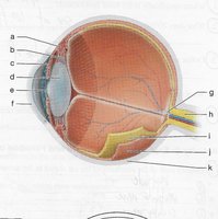
Key Terms:
Cornea, iris, pupil, lens, retina, optic nerve, optic disk, suspensory ligaments, sclera, choroid, ciliary body
Step-by-Step Guidance
Study the diagram and locate each label (a–k).
Use your lab manual to match each label to the correct anatomical structure.
Pay attention to the unique features of each part (e.g., the colored part is the iris, the hole is the pupil).
Write the correct anatomical term for each label, but stop before completing all labels.
Try solving on your own before revealing the answer!
Lab Manual Unit 17: Labeling – Ear
Background
Topic: Anatomy of the Human Ear
This section asks you to identify the labeled parts of the human ear in a diagram. Understanding the anatomy of the ear is important for learning about hearing and balance.
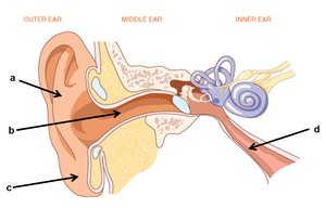
Key Terms:
Outer ear, middle ear, inner ear, auditory canal, tympanic membrane, cochlea, vestibule, semicircular canals
Step-by-Step Guidance
Study the diagram and locate each label (a–d).
Use your lab manual to match each label to the correct anatomical structure.
Pay attention to the location and shape of each part (e.g., the outer ear, the canal, the cochlea).
Write the correct anatomical term for each label, but stop before completing all labels.
Try solving on your own before revealing the answer!
Lab Manual Unit 17: Labeling – Middle and Inner Ear
Background
Topic: Anatomy of the Middle and Inner Ear
This section asks you to identify the labeled parts of the middle and inner ear in a diagram. This is important for understanding hearing and balance, as well as the role of cranial nerves.
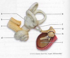
Key Terms:
Vestibule, cochlear branch, vestibular branch, auditory ossicles, semicircular canals, cranial nerve VIII
Step-by-Step Guidance
Study the diagram and locate each label (a–i).
Use your lab manual to match each label to the correct anatomical structure.
Pay attention to the function of each part (e.g., balance, hearing, nerve branches).
Write the correct anatomical term for each label, but stop before completing all labels.
Try solving on your own before revealing the answer!
