 Back
Language of Anatomy: Anatomical Terms, Positions, Planes, and Body Cavities
Study Guide - Smart Notes
 Tailored notes based on your materials, expanded with key definitions, examples, and context.
Language of Anatomy
Studying Anatomy and Anatomical Terms
Anatomy uses a specialized language derived from Greek, Latin, Italian, and sometimes Englishto describe locations, regions, and relationships within the human body. Understanding these terms is essential for clear communication among healthcare professionals.
Anatomical terms are standardized to avoid confusion.
Names often reflect the shape, location, or function of the structure.
Body Regions and Anatomical Landmarks
The human body is divided into regions for descriptive purposes. These regions are identified using specific anatomical terms.
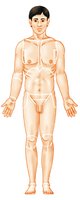
Anterior (front) regions include nasal (nose), oral (mouth), cervical (neck), frontal (forehead), orbital (eye), buccal (cheek), mental (chin), acromial (shoulder), axillary (armpit), brachial (arm), antecubital (front of elbow), abdominal (abdomen), pelvic (pelvis), antebrachial (forearm), carpal (wrist), palmar (palm), pollex (thumb), digital (fingers), mammary (breast), sternal (breastbone), thoracic (chest), umbilical (navel), pubic (genital region), patellar (anterior knee), crural (leg), tarsal (ankle), pedal (foot), digital (toes), inguinal (groin), coxal (hip), femoral (thigh), fibular/peroneal (side of leg), hallux (great toe).
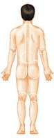
Posterior (back) regions include otic (ear), occipital (back of head), cephalic (head), vertebral (spinal column), scapular (shoulder blade), dorsum/dorsal (back), olecranal (back of elbow), lumbar (loin), sacral (between hips), gluteal (buttock), perineal (region between anus and external genitalia), femoral (thigh), popliteal (back of knee), sural (calf), calcaneal (heel), plantar (sole), manus (hand).
Axial and Appendicular Divisions
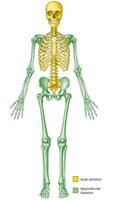
The body is divided into two main regions: axial and appendicular. The axial region includes the head, neck, and trunk, while the appendicular region consists of the limbs.
Axial skeleton: Skull, vertebral column, and rib cage.
Appendicular skeleton: Limbs and girdles (shoulder and pelvic).
Anatomical Position
Definition and Importance
The anatomical position is a standardized posture used as a reference in anatomy. The body is erect, feet slightly apart, palms facing forward, and thumbs pointing away from the body. This position ensures consistency in describing locations and directions.
All directional terms are based on the anatomical position.
It is essential for accurate communication in medical settings.

Supination and Pronation
Definitions and Applications
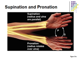
Supination and pronation describe movements of the forearm. Supination is the rotation that turns the palm upward, while pronation turns the palm downward.
Supination: Radius and ulna are parallel.
Pronation: Radius rotates over the ulna.
Directional Terms
Commonly Used Directional Terms
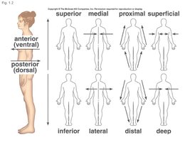
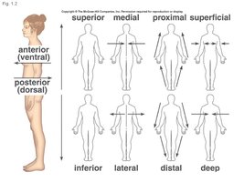
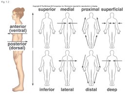
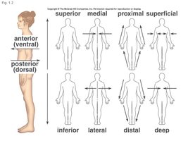
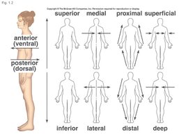
Directional terms are used to describe the locations of structures relative to other structures or locations in the body. These terms are often paired with their opposites.
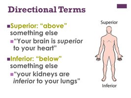
Superior (cranial): Toward the head or upper part of a structure.
Inferior (caudal): Away from the head or toward the lower part.
Anterior (ventral): Toward the front of the body.
Posterior (dorsal): Toward the back of the body.
Medial: Toward the midline of the body.
Lateral: Away from the midline.
Intermediate: Between medial and lateral.
Proximal: Closer to the origin of a body part or point of attachment.
Distal: Farther from the origin or point of attachment.
Superficial (external): Toward or at the body surface.
Deep (internal): Away from the body surface.
Right and Left
In anatomy, right and left always refer to the patient's right and left, not the observer's. This convention is critical to avoid errors in medical practice.
Always use the patient's perspective when describing right and left.
Planes of Dissection
Body Planes
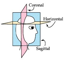
Body planes are imaginary lines used to divide the body for anatomical study. They correspond to the x, y, and z axes.
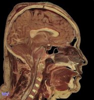
Sagittal plane: Divides the body into right and left parts.
Midsagittal (median) plane: Lies on the midline.
Parasagittal plane: Not on the midline.
Frontal (coronal) plane: Divides the body into anterior and posterior parts.
Transverse (horizontal) plane: Divides the body into superior and inferior parts.
Oblique section: Cuts made diagonally, typically avoided for clarity.
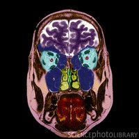
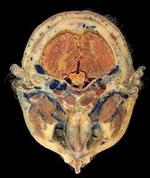
Planes Through an Organ or Tube
Longitudinal: Cut along the length of an organ.
Transverse (cross): Cut at a right angle to the length.
Oblique: Cut at any angle other than a right angle.
Body Cavities
Classification of Body Cavities
Body cavities are spaces within the body that house internal organs. They are classified as enclosed (do not open to the outside) or exposed (open to the exterior).
Enclosed cavities: Require penetration of the body wall for access.
Exposed cavities: Can be accessed without penetrating the body wall.
Dorsal and Ventral Body Cavities
The two main body cavities are the dorsal and ventral cavities.
Dorsal cavity: Protects the nervous system; divided into cranial (brain) and vertebral (spinal cord) cavities.
Ventral cavity: Contains internal organs; subdivided into thoracic (heart and lungs) and abdominopelvic (digestive, urinary, and reproductive organs) cavities.
Thoracic and Abdominopelvic Cavities
Thoracic cavity: Subdivided into pleural cavities (lungs), mediastinum (central region), and pericardial cavity (heart).
Abdominopelvic cavity: Separated from thoracic cavity by the diaphragm; includes abdominal (digestive organs) and pelvic (bladder, reproductive organs, rectum) cavities.
Serous Membranes
Serous membranes (serosa) line the ventral body cavities. They consist of two layers:
Parietal serosa: Adheres to the cavity wall.
Visceral serosa: Adheres to the organ.
The cavity between the layers contains serous fluid, which lubricates and reduces friction.
Serous Membrane Relationships
Pericardium: Surrounds the heart.
Pleura: Surrounds the lungs.
Peritoneum: Surrounds abdominopelvic organs.
Other Body Cavities
Oral and digestive: Mouth and digestive organ cavities.
Nasal: Within and posterior to the nose.
Orbital: Houses the eyes.
Middle ear: Contains ossicles for sound transmission.
Synovial: Joint cavities.
Abdominopelvic Quadrants and Regions
Quadrants
The abdominopelvic cavity is divided into four quadrants for clinical reference: right upper, left upper, right lower, and left lower. These divisions are made by a vertical and horizontal line through the umbilicus (navel).
Regions
Anatomists use nine regions, similar to a tic-tac-toe board, to describe locations within the abdominopelvic cavity. These regions are delineated by four planes.
Right hypochondriac, epigastric, left hypochondriac
Right lumbar, umbilical, left lumbar
Right iliac (inguinal), hypogastric (pubic), left iliac (inguinal)
Each region contains specific organs, aiding in diagnosis and communication.
Region | Organs |
|---|---|
Right hypochondriac | Liver, gallbladder |
Epigastric | Stomach |
Left hypochondriac | Diaphragm, spleen |
Right lumbar | Ascending colon |
Umbilical | Small intestine, transverse colon |
Left lumbar | Descending colon |
Right iliac | Cecum, appendix |
Hypogastric | Urinary bladder |
Left iliac | Initial part of sigmoid colon |
Summary
Understanding anatomical terminology, positions, planes, and body cavities is foundational for studying human anatomy and communicating effectively in healthcare settings. Mastery of these concepts enables precise description and localization of structures and processes within the body.
