 Back
BackLanguage of Anatomy: Foundational Concepts and Terminology
Study Guide - Smart Notes
 Tailored notes based on your materials, expanded with key definitions, examples, and context.
Language of Anatomy
Studying Anatomy
Anatomy is the study of the structure of the human body. To facilitate clear communication, anatomists use a specialized language derived from Greek, Latin, Italian, and sometimes English. This terminology allows precise identification and description of body parts and their relationships.
Anatomical Terms: Standardized names for body regions and structures.
Importance: Reduces confusion and errors in medical and scientific settings.
Example: The term cervical refers to the neck region.
Anatomical Position and Body Regions
Anatomical Position
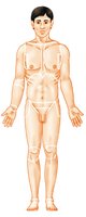
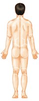
The anatomical position is a reference posture used to describe locations and relationships of body parts. The body is erect, feet slightly apart, palms facing forward, and thumbs pointing away from the body. This position standardizes descriptions and is essential for anatomical terminology.
Body Regions: Specific areas of the body are named for clarity (e.g., nasal for nose, oral for mouth).
Anterior (front) and Posterior (back) Views: Used to describe the location of structures.
Axial and Appendicular Divisions
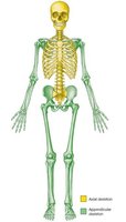
The human body is divided into two main regions: the axial and appendicular divisions. The axial division includes the head, neck, and trunk, while the appendicular division consists of the limbs.
Axial: Head, neck, trunk
Appendicular: Arms and legs
Directional Terms
Common Directional Terms
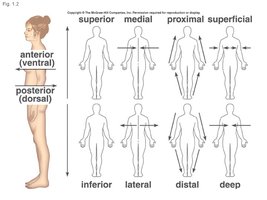
Directional terms are used to describe the locations of structures relative to other structures or locations in the body. These terms are often paired with their opposites.
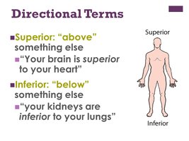
Superior (cranial): Toward the head or upper part of a structure
Inferior (caudal): Away from the head or toward the lower part
Anterior (ventral): Toward the front of the body
Posterior (dorsal): Toward the back of the body
Medial: Toward the midline
Lateral: Away from the midline
Proximal: Closer to the point of attachment
Distal: Farther from the point of attachment
Superficial (external): Toward or at the body surface
Deep (internal): Away from the body surface
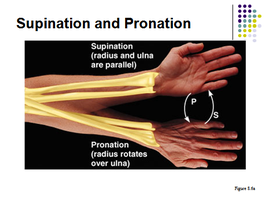
Supination vs. Pronation
Supination and pronation describe movements of the forearm. Supination is the rotation that turns the palm upward, while pronation turns the palm downward.
Supination: Radius and ulna are parallel
Pronation: Radius rotates over ulna
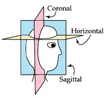
Planes of Dissection
Body Planes
Body planes are imaginary lines used to divide the body for anatomical study. These planes correspond to the x, y, and z axes and are essential for understanding sectional anatomy.
Sagittal Plane: Divides the body into right and left parts
Midsagittal (Median) Plane: Lies on the midline
Parasagittal Plane: Not on the midline
Frontal (Coronal) Plane: Divides the body into anterior and posterior parts
Transverse (Horizontal) Plane: Divides the body into superior and inferior parts
Oblique Plane: Cuts made diagonally
Body Cavities and Membranes
Body Cavities
Body cavities are spaces within the body that house internal organs. They are classified as dorsal (posterior) and ventral (anterior) cavities.
Dorsal Cavity: Protects the nervous system; includes cranial and vertebral cavities
Ventral Cavity: Contains internal organs; subdivided into thoracic and abdominopelvic cavities
Thoracic Cavity: Contains heart and lungs
Abdominopelvic Cavity: Contains digestive organs, bladder, reproductive organs
Serous Membranes
Serous membranes (serosa) line the ventral body cavities and cover the organs within these cavities. They consist of two layers: parietal (lines cavity wall) and visceral (covers organ). The space between these layers contains serous fluid, which reduces friction.
Pericardium: Surrounds the heart
Pleura: Surrounds the lungs
Peritoneum: Surrounds abdominopelvic organs
Abdominopelvic Regions and Quadrants
Abdominopelvic Regions
The abdominopelvic area is divided into nine regions for anatomical study and four quadrants for clinical use. These divisions help localize pain, injuries, or disease.
Nine Regions: Right/left hypochondriac, epigastric, right/left lumbar, umbilical, right/left iliac (inguinal), hypogastric (pubic)
Four Quadrants: Right upper, left upper, right lower, left lower
Summary Table: Directional Terms
Term | Definition | Example |
|---|---|---|
Superior | Above another structure | The brain is superior to the heart |
Inferior | Below another structure | The kidneys are inferior to the lungs |
Anterior | Toward the front | The sternum is anterior to the heart |
Posterior | Toward the back | The vertebral column is posterior to the heart |
Medial | Toward the midline | The nose is medial to the eyes |
Lateral | Away from the midline | The ears are lateral to the nose |
Proximal | Closer to point of attachment | The elbow is proximal to the wrist |
Distal | Farther from point of attachment | The fingers are distal to the elbow |
Superficial | Toward the surface | The skin is superficial to muscles |
Deep | Away from the surface | The bones are deep to the skin |
