 Back
BackLeukocytes and the Immune System: Structure, Function, and Defense Mechanisms
Study Guide - Smart Notes
 Tailored notes based on your materials, expanded with key definitions, examples, and context.
Leukocytes: Structure, Types, and Functions
Overview of Leukocytes
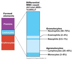
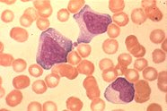
Leukocytes, or white blood cells (WBCs), are the only formed elements in blood that are complete cells. They play a critical role in defending the body against bacteria, viruses, parasites, toxins, and tumor cells. Leukocytes can leave the bloodstream (diapedesis) to mount immune and inflammatory responses, move via amoeboid motion, and are attracted to sites of infection or injury by chemotaxis.
Normal count: 4,000–11,000 WBCs/μl (less than 1% of total blood volume)
Leukocytosis: WBC count >11,000/μl, often in response to infection
Classification: Granulocytes and Agranulocytes
Types of Leukocytes
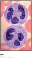
Neutrophils: >50% of leukocytes; phagocytes that ingest and destroy bacteria, especially during acute infections. Cytoplasm contains granules with hydrolytic enzymes and defensins.
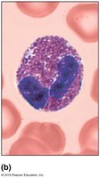
Eosinophils: 2–4% of leukocytes; combat parasitic worms by releasing digestive enzymes and modulate immune responses, especially in allergies and asthma.
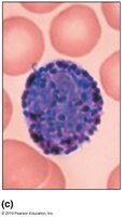
Basophils: Rarest WBCs (0.5%); granules contain histamine, a vasodilator that attracts other WBCs and increases blood vessel permeability.
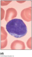
Lymphocytes: 25–45% of leukocytes; large nucleus, found in lymphoid tissues. T cells act directly against infected or cancerous cells; B cells produce antibodies.
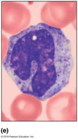
Monocytes: 3–8% of leukocytes; largest WBCs, differentiate into macrophages in tissues, phagocytic in chronic infections.
Granulocytes vs. Agranulocytes
Granulocytes: Neutrophils, eosinophils, basophils; have visible cytoplasmic granules and lobed nuclei.
Agranulocytes: Lymphocytes, monocytes; lack visible granules, nuclei are spherical or kidney-shaped.
Leukopoiesis: Formation of Leukocytes
Process of Leukopoiesis
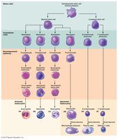
Leukopoiesis is the process of leukocyte production, regulated by hormones such as interleukins and colony-stimulating factors. It begins with a hemocytoblast (stem cell) that differentiates into myeloid or lymphoid stem cells, which then give rise to various leukocyte lineages.
Myeloid stem cells: Produce granulocytes (neutrophils, eosinophils, basophils) and monocytes.
Lymphoid stem cells: Produce lymphocytes (T and B cells).
Lifespan: Granulocytes live 0.25–9 days; most die fighting infections.
Leukocyte Disorders
Major Disorders
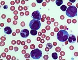
Leukemia: Cancer of WBCs, often due to uncontrolled proliferation of unspecialized cells. Leads to anemia, clotting problems, and increased infection risk. Treatment includes chemotherapy, irradiation, and bone marrow transplant.
Infectious Mononucleosis: Caused by Epstein-Barr virus; characterized by excessive, abnormal lymphocytes, fatigue, sore throat, and fever.
Leukopenia: Abnormally low WBC count, often due to drugs such as glucocorticoids or chemotherapy agents.
The Immune System: Innate and Adaptive Defenses
Overview of Immune Defenses
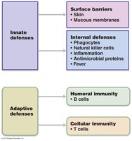
The immune system is divided into innate (nonspecific) and adaptive (specific) defenses. Innate defenses provide immediate, general protection, while adaptive defenses target specific pathogens and involve immunological memory.
Innate defenses: Surface barriers (skin, mucous membranes), phagocytes, natural killer cells, inflammation, antimicrobial proteins, fever.
Adaptive defenses: Humoral immunity (B cells/antibodies), cellular immunity (T cells).
First Line of Defense: Surface Barriers
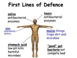
Skin and mucous membranes form the first line of defense, providing physical and chemical barriers to infection.
Acidity: Skin, vaginal, and stomach secretions deter bacterial growth.
Enzymes: Lysozyme in saliva, mucus, and tears; protein-digesting enzymes in the stomach.
Mucin: Traps microorganisms in digestive and respiratory tracts.
Defensins: Antimicrobial peptides secreted by skin and mucous membranes.
Other chemicals: Sebum and dermicidin are toxic to bacteria.
Innate Immune Activation: PAMPs and DAMPs
Recognition of Danger Signals
The innate immune system is activated by PAMPs (Pathogen-Associated Molecular Patterns) and DAMPs (Damage-Associated Molecular Patterns). PAMPs are found on pathogens, while DAMPs are endogenous molecules released by damaged cells. Recognition occurs via pattern recognition receptors such as Toll-like receptors, leading to phagocytosis, inflammation, and activation of the adaptive immune system.
Inflammation: Mechanisms and Benefits
Inflammatory Response
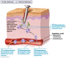
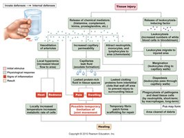
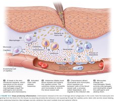
Inflammation is a protective response to injury or infection, characterized by redness, heat, swelling, and pain. It prevents the spread of pathogens, disposes of debris, initiates repair, and alerts the adaptive immune system. Inflammatory chemicals (e.g., histamine, kinins, prostaglandins, complement) increase blood flow and vessel permeability, attracting immune cells to the site.
Margination: Leukocytes cling to capillary walls.
Diapedesis: Leukocytes squeeze through capillary walls into tissues.
Chemotaxis: Leukocytes follow chemical signals to the site of injury.
Antimicrobial Proteins: Interferons and Complement
Interferons
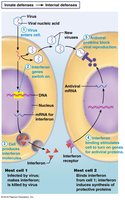
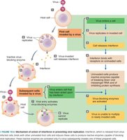
Interferons are proteins secreted by virus-infected cells. They protect neighboring cells by blocking viral protein synthesis and degrading viral RNA. Interferons also activate macrophages and natural killer cells, contributing to anti-cancer defenses.
Complement System
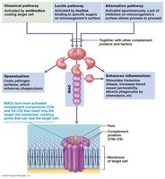
The complement system consists of at least 20 plasma proteins that circulate in an inactive state. Once activated, they amplify inflammation, promote phagocytosis (opsonization), and directly lyse pathogens via the membrane attack complex (MAC).
Fever: Systemic Response to Infection
Mechanism and Effects of Fever
Fever is an elevation of body temperature, triggered by pyrogens released from leukocytes and macrophages in response to infection. Moderate fever enhances immune function by increasing metabolic rate and sequestering iron and zinc, which bacteria need to multiply. High fever can be dangerous due to protein denaturation and cellular dysfunction.

Natural Killer (NK) Cells
Role and Activation of NK Cells
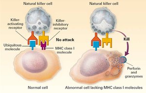
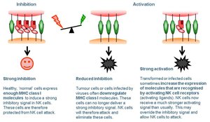
Natural killer cells are lymphocytes that provide rapid, nonspecific defense against virus-infected and cancerous cells. They recognize cells with reduced MHC I expression and/or stress markers, releasing perforin and granzymes to induce apoptosis. NK cells also express Toll-like receptors, allowing them to respond to PAMPs and DAMPs.
Summary Table: Leukocyte Types and Functions
Leukocyte Type | Structure | Function | Relative Abundance (%) |
|---|---|---|---|
Neutrophil | Multi-lobed nucleus, pale granules | Phagocytosis of bacteria | 50–70 |
Eosinophil | Bilobed nucleus, red granules | Combat parasitic worms, modulate allergies | 2–4 |
Basophil | S- or U-shaped nucleus, dark granules | Release histamine, mediate inflammation | 0.5–1 |
Lymphocyte | Large nucleus, thin rim of cytoplasm | Adaptive immunity (B and T cells) | 25–45 |
Monocyte | Kidney-shaped nucleus, abundant cytoplasm | Differentiate into macrophages, phagocytosis | 3–8 |
