 Back
BackLeukocytes: Structure, Function, and Classification
Study Guide - Smart Notes
 Tailored notes based on your materials, expanded with key definitions, examples, and context.
Leukocytes (White Blood Cells)
Overview of Leukocytes
Leukocytes, or white blood cells (WBCs), are essential components of the immune system, responsible for defending the body against pathogens and foreign substances. They are larger than erythrocytes (red blood cells) and possess prominent nuclei.
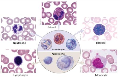
Leukocytes are classified into two main groups: Granulocytes and Agranulocytes.
Granulocytes contain cytoplasmic granules that release substances when activated, while agranulocytes lack these granules.
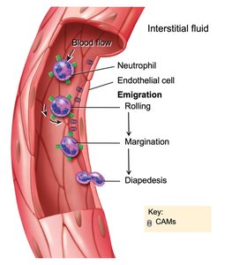
Leukocytes are involved in processes such as chemotaxis, margination, and diapedesis, which enable them to migrate to sites of infection or injury.
Functional Characteristics of Leukocytes
Leukocytes exhibit specialized behaviors that allow them to respond to infection and inflammation.
Chemotaxis: Attraction to chemical stimuli released by damaged tissues or pathogens, guiding leukocytes to the site of infection.
Margination: The process by which leukocytes adhere to the endothelial cells of blood vessels via cell adhesion molecules (CAMs), positioning them for migration.
Diapedesis: The ability of leukocytes to squeeze between endothelial cells and exit the bloodstream, entering tissues to combat pathogens.
Classification of Leukocytes
Granulocytes
Granulocytes are characterized by the presence of granules in their cytoplasm, which stain with specific dyes.
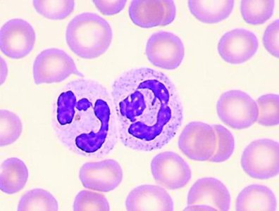
Neutrophils: Most abundant WBC (50–70%), polymorphonuclear (3–5 lobes), stains with both acidic and basic dyes.
Phagocytic; digest bacteria using lysosomal enzymes.
Produce hydrogen peroxide during the respiratory burst to kill pathogens.
First responders to infection due to their high numbers.
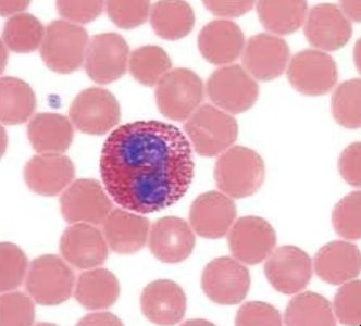
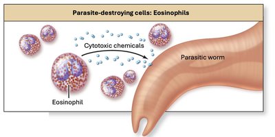
Eosinophils: Bi-lobed nucleus, stains with acidic dyes (pink granules), 1–4% of WBCs.
Attack large parasites, especially in blood and mucosal linings.
Increase in allergic reactions and asthma.
Produce histaminases to decrease inflammation.
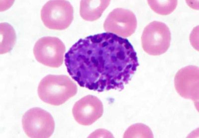
Basophils: Least abundant (0.5–1%), stains blue/purple with basic dyes, S-shaped nucleus often hard to see.
Granules release histamine (vasodilator), heparin (anticoagulant), and eicosanoids (increase inflammation).
Enhance effects of mast cells and initiate inflammation.
Agranulocytes
Agranulocytes lack visible granules in their cytoplasm and have more prominent nuclei.
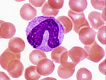
Monocytes: Largest WBC (3–8%), leave blood to become macrophages.
Elevated in chronic infections.
Phagocytize pathogens and present antigens to lymphocytes.
Attract fibroblasts to create scar tissue during healing.
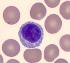
Lymphocytes: 20–40% of WBCs, three classes:
T Cells: Cellular immunity.
B Cells: Antibody-mediated immunity.
Natural Killer (NK) Cells: Immunological surveillance.
Leukocyte Profiles and Cell Numbers
Differential Count and Clinical Significance
The number and proportion of each type of leukocyte can change in response to various conditions, making cell counts clinically important.
Differential count: Measures the number of each type of WBC in a blood sample.
Leukopenia: Inadequate numbers of WBCs, impairing immune response.
Leukocytosis: Excessive numbers of WBCs, often normal during infection but may indicate disease if persistent.
High numbers of a particular cell type may indicate specific illnesses.
Leukopoiesis: Formation of Leukocytes
Hematopoietic Stem Cells and Lineages
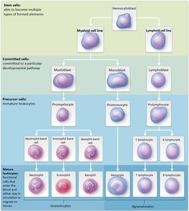
Leukopoiesis is the process of leukocyte formation from hematopoietic stem cells (hemocytoblasts) in the bone marrow.
Hemocytoblast: Stem cell capable of forming any blood cell (WBC, RBC, platelets).
Two main cell lines:
Myeloid line: Forms granulocytes and monocytes.
Lymphoid line: Forms lymphocytes.
Committed cells include myeloblasts (granulocytes), monoblasts (monocytes), and lymphoblasts (lymphocytes).
The type of cell produced in excess can indicate specific illnesses.
Summary Table: Leukocyte Types and Functions
Type | Appearance | Function | Normal % |
|---|---|---|---|
Neutrophil | 3–5 lobed nucleus, pale granules | Phagocytosis, first responder to infection | 50–70% |
Eosinophil | Bi-lobed nucleus, pink granules | Attack parasites, modulate allergic responses | 1–4% |
Basophil | S-shaped nucleus, blue/purple granules | Release histamine, heparin, inflammation | 0.5–1% |
Monocyte | Large, kidney-shaped nucleus | Phagocytosis, antigen presentation, chronic infection | 3–8% |
Lymphocyte | Large, round nucleus | T, B, NK cell immunity | 20–40% |
Key Terms and Concepts
Phagocytosis: Cellular process of engulfing and digesting pathogens.
Respiratory burst: Production of reactive oxygen species (e.g., hydrogen peroxide) to kill microbes.
Histaminase: Enzyme that breaks down histamine, reducing inflammation.
Antigen presentation: Display of pathogen fragments to lymphocytes to initiate immune response.
Immunological surveillance: Monitoring for abnormal cells by NK cells.
Equations and Formulas
Leukocyte percentage calculation:
Clinical Application Example
Example: A patient with elevated eosinophil count may be experiencing a parasitic infection or allergic reaction.
Example: Leukopenia can result from chemotherapy, increasing susceptibility to infection.
