 Back
BackLife-Span Development of the Brain and Behavior: Neural Development and Plasticity
Study Guide - Smart Notes
 Tailored notes based on your materials, expanded with key definitions, examples, and context.
Neural Development
Introduction to Neural Development
The development of the nervous system is a complex, multi-stage process that transforms a fertilized egg into a highly organized brain and spinal cord. This process involves the coordinated proliferation, migration, differentiation, and connection of billions of neurons and glial cells.
Embryonic Origins of the Nervous System
Zygote: The fertilized egg that begins the process of development.
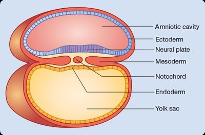
After one week, the embryo forms three primary germ layers:
Endoderm: Inner layer
Mesoderm: Middle layer
Ectoderm: Outer layer; gives rise to the nervous system
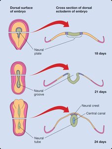
The neural plate forms from the ectoderm, which folds to create the neural groove and eventually the neural tube.
The neural tube develops into the central nervous system (CNS).

Major Brain Regions in Development
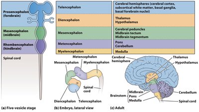
The embryonic brain forms three primary vesicles, which further subdivide into five secondary vesicles, giving rise to the adult brain structures:
Prosencephalon (forebrain)
Mesencephalon (midbrain)
Rhombencephalon (hindbrain)
Genetic and Environmental Influences
Genotype: The genetic makeup of an individual.
Phenotype: The observable characteristics, which can change throughout life due to environmental influences (phenotypic plasticity).
Gene expression: The process by which genes are transcribed and translated into proteins.
Genetic predisposition: Increased likelihood of developing certain traits or diseases due to inherited genes.
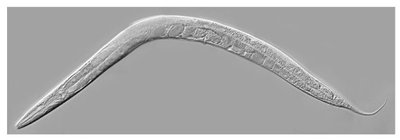
Development in invertebrates (e.g., C. elegans) is highly predetermined, while vertebrate development is shaped by cell-cell interactions and environmental factors.
Stages of Nervous System Development
Overview of Developmental Stages
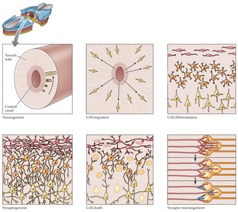
Neural development proceeds through six major stages:
Neurogenesis
Cell migration
Differentiation
Synaptogenesis
Neuronal cell death (apoptosis)
Synapse rearrangement
1. Neurogenesis
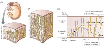
Neurogenesis is the rapid proliferation of nerve cells after the neural tube forms. Non-neural cells in the ventricular zone divide by mitosis, producing neurons and glial cells.
During peak proliferation, up to 250,000 new neurons are generated per minute.
2. Cell Migration
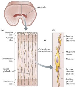
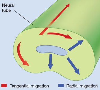
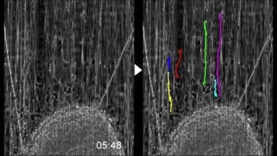
Newly formed cells migrate from the ventricular zone to their final destinations in the brain. Migration occurs in two main patterns:
Radial migration: Guided by radial glial cells, moving outward from the ventricular zone.
Tangential migration: Movement parallel to the brain surface, often used by interneurons.
Cell adhesion molecules (CAMs) and chemotrophic factors guide migration.
3. Differentiation
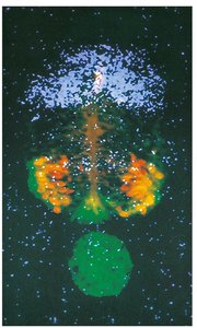
Once cells reach their destinations, they differentiate into specific types (e.g., neurons, glia) by expressing unique sets of genes. Differentiation is influenced by:
Induction: Neighboring cells release signals (e.g., Sonic hedgehog from the notochord) that direct cell fate.
Regulation: If cells are lost, undifferentiated stem cells can replace them.
4. Synaptogenesis
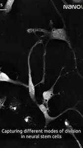
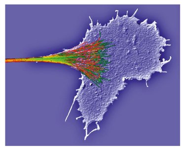
Synaptogenesis is the formation of synapses between neurons. Growth cones at the tips of axons and dendrites extend filopodia to find target cells, guided by CAMs, chemoattractants, and chemorepellents.
Growth cones are dynamic structures that explore the environment and form connections.
Trophic factors (e.g., nerve growth factor, brain-derived neurotrophic factor) support axon growth and synapse formation.
5. Neuronal Cell Death (Apoptosis)
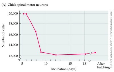
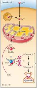
Programmed cell death (apoptosis) is a normal part of development, removing excess neurons. Apoptosis is regulated by:
Death genes (e.g., caspases) activated only during apoptosis.
Neurotrophic factors: Neurons compete for limited amounts; those that do not receive enough undergo apoptosis.
Bcl-2 proteins block apoptosis by preventing release of pro-apoptotic factors.
6. Synapse Rearrangement
After initial synapse formation, many synapses are retracted or reorganized based on activity and experience. Active synapses are stabilized, while inactive ones are eliminated.
Experience and neural activity shape the final pattern of connectivity.
Postnatal Brain Development and Plasticity
Postnatal Changes in the Brain
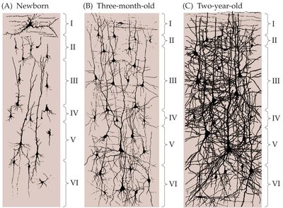
Brain volume increases fourfold from birth to adulthood, mainly due to synaptogenesis, dendritic branching, and myelination.
Myelination of axons allows for faster neural communication and continues into young adulthood.
Pruning of synapses and grey matter occurs during adolescence, especially in the prefrontal cortex.
Sensitive and Critical Periods
There are specific windows during development when the brain is especially sensitive to environmental input:
Sensitive periods: Times when experience has a strong effect on development, but some plasticity remains afterward.
Critical periods: Essential windows when certain experiences must occur for normal development (e.g., vision, language acquisition).
Neural Plasticity and Reorganization
The brain can reorganize after injury or sensory deprivation, especially during early development.
Plasticity is supported by synaptic remodeling, dendritic growth, and, in some regions, adult neurogenesis (e.g., hippocampus).
Neurodegeneration and Aging
Neurodegenerative Diseases
Dementia: Severe cognitive decline, including memory loss.
Alzheimer’s disease: Characterized by β-amyloid plaques, neurofibrillary tangles (Tau protein), and loss of cholinergic neurons.
Diagnosis can involve PET scans and blood tests for β-amyloid.
Adult Neurogenesis and Environmental Effects
New neurons are generated in the adult hippocampus, olfactory bulbs, and striatum.
Enriched environments, physical activity, and intellectual engagement promote dendritic growth and synaptic complexity.
Summary Table: Stages of Nervous System Development
Stage | Description | Key Mechanisms |
|---|---|---|
Neurogenesis | Birth of new neurons from progenitor cells | Mitosis in ventricular zone |
Cell Migration | Movement of neurons to final positions | Radial glia, CAMs, chemotrophic factors |
Differentiation | Specialization into neuron/glia types | Induction, gene expression |
Synaptogenesis | Formation of synaptic connections | Growth cones, CAMs, trophic factors |
Neuronal Cell Death | Programmed elimination of excess neurons | Apoptosis, neurotrophic factor competition |
Synapse Rearrangement | Refinement of synaptic connections | Activity-dependent pruning |
Example: If a child is deprived of visual input during the critical period, the visual cortex will not develop normal connections, resulting in permanent deficits.
Additional info: Adult neurogenesis is an active area of research, with implications for learning, memory, and recovery from brain injury.
