 Back
Liver, Gallbladder, and Pancreas: Anatomy and Physiology Study Guide
Study Guide - Smart Notes
 Tailored notes based on your materials, expanded with key definitions, examples, and context.
Liver, Gallbladder, and Pancreas
Overview of Accessory Digestive Organs
The liver, gallbladder, and pancreas are essential accessory organs that support the digestive functions of the small intestine. Each organ has specialized roles in digestion, absorption, and metabolic regulation.
Liver: Produces bile, a fat emulsifier necessary for digestion.
Gallbladder: Stores and releases bile as needed.
Pancreas: Supplies digestive enzymes and bicarbonate to neutralize stomach acid.
The Pancreas
Gross Anatomy and Location
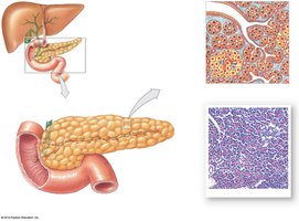
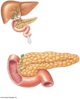
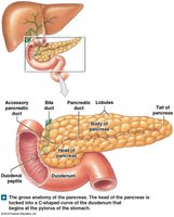
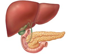
The pancreas is a retroperitoneal organ located deep to the greater curvature of the stomach. Its head is encircled by the duodenum, and its tail abuts the spleen.
Head: Located within the C-shaped curve of the duodenum.
Body and Tail: Extend toward the spleen.
Cellular Organization
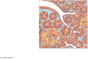
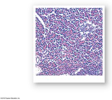
The pancreas contains both exocrine and endocrine cells:
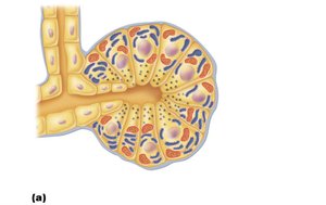
Exocrine cells (acini): Produce digestive enzymes and secrete them into ducts.
Endocrine cells (islets): Secrete hormones such as insulin and glucagon.
Exocrine Function and Pancreatic Juice
The pancreas produces 1200–1500 mL of pancreatic juice daily, which is a watery, alkaline solution (pH 8) containing:
Electrolytes: Primarily bicarbonate (HCO3-).
Digestive enzymes:
Proteases: Digest proteins; secreted in inactive form.
Amylase: Digests starch.
Lipase: Digests triglycerides.
Nucleases: Digest DNA and RNA.
Activation of Pancreatic Proteases
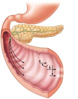
Proteases are secreted in inactive forms to prevent self-digestion. Activation occurs in the duodenum:
Enteropeptidase: Activates trypsinogen to trypsin.
Trypsin: Activates more trypsinogen, procarboxypeptidase, and chymotrypsinogen.
Endocrine Function
The endocrine pancreas consists of islet cells that secrete:
Insulin: Lowers blood glucose.
Glucagon: Raises blood glucose.
The Liver
Gross Anatomy
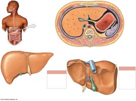
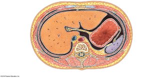
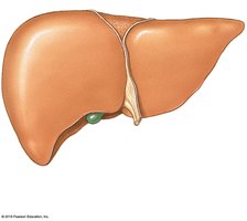
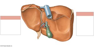
The liver is the largest internal organ (~3 lbs), consisting of four primary lobes: right, left, caudate, and quadrate.
Falciform ligament: Separates right and left lobes; suspends liver from diaphragm and anterior abdominal wall.
Round ligament (ligamentum teres): Remnant of fetal umbilical vein.
Blood Flow Through the Hepatic Portal System
The hepatic portal system delivers nutrient-rich blood from the digestive tract to the liver for processing and detoxification. 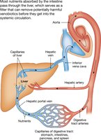
Microscopic Anatomy: Liver Lobules
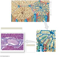
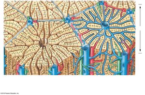
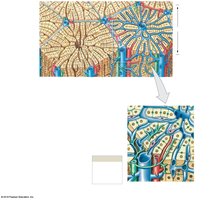
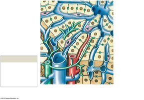
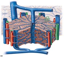
Liver lobules are hexagonal units composed of plates of hepatocytes. Each lobule contains:
Central vein: Located in the longitudinal axis.
Portal triad: At each corner, consisting of a branch of the hepatic artery, hepatic portal vein, and bile duct.
Hepatocyte Functions
Hepatocytes are highly metabolic cells with abundant rough and smooth ER, Golgi apparatus, peroxisomes, and mitochondria. Their functions include:
Bile production: ~900 mL per day.
Metabolic regulation: Processing nutrients, storing glucose as glycogen, synthesizing plasma proteins.
Vitamin storage: Fat-soluble vitamins (D, E, A, K).
Detoxification: Converting ammonia to urea.
Bile: Composition and Enterohepatic Circulation
Bile is a yellow-green, alkaline solution containing:
Bile salts: Cholesterol derivatives for fat emulsification and absorption.
Bilirubin: Pigment from heme breakdown; converted to stercobilin in intestine (brown color of feces).
Cholesterol, triglycerides, phospholipids, electrolytes.
Enterohepatic Circulation
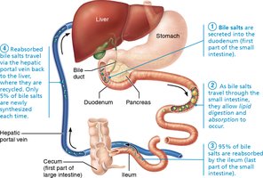
Bile salts are recycled via the enterohepatic circulation:
Reabsorbed in the ileum.
Returned to the liver via hepatic portal blood.
Re-secreted in newly formed bile.
About 95% of bile salts are recycled.
The Gallbladder
Anatomy and Function
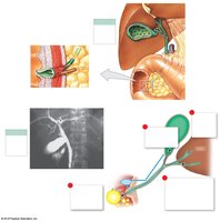
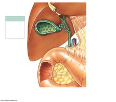
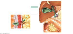
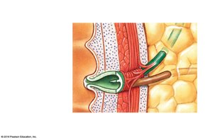
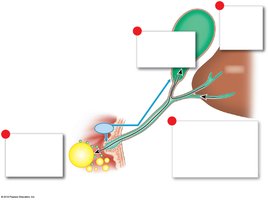
The gallbladder is a thin-walled muscular sac on the ventral surface of the liver. Its functions include:
Storage and concentration of bile: Absorbs water and ions.
Release of bile: Muscular contractions expel bile via the cystic duct into the bile duct.
Gallbladder Physiology
Bile is secreted continuously by the liver (~1 L/day).
Bile becomes more concentrated in the gallbladder.
Release of cholecystokinin (CCK) triggers contraction of the gallbladder and relaxation of the hepatopancreatic sphincter, ejecting bile into the duodenum.
Bile salts emulsify lipids in the digestive tract.
Homeostatic Imbalance: Gallstones and Jaundice
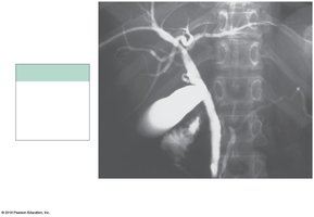
Gallstones (biliary calculi): Caused by excess cholesterol or insufficient bile salts; can obstruct bile flow and cause pain.
Obstructive jaundice: Blockage leads to accumulation of bile salts and pigments in blood, resulting in yellow skin.
Treatment: Includes drugs, ultrasound (lithotripsy), laser vaporization, or surgery.
Small Intestine
Gross Anatomy and Subdivisions
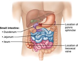
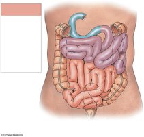
The small intestine is the major organ of digestion and absorption, measuring 2–4 meters in length.
Duodenum: ~25 cm, curves around the pancreas.
Jejunum: ~2.5 m, attached by mesentery.
Ileum: ~3.6 m, joins the large intestine at the ileocecal valve.
Microscopic Anatomy: Structural Modifications for Absorption
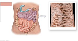
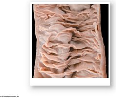
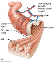
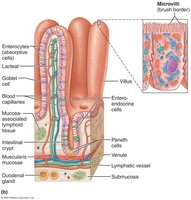
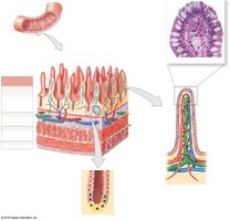
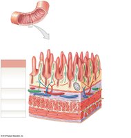
The small intestine has several modifications to increase surface area for absorption:
Circular folds: Permanent folds (~1 cm deep) that slow chyme movement.
Villi: Fingerlike projections (~1 mm high) with capillary beds and lacteals for nutrient absorption.
Microvilli: Cytoplasmic extensions forming the brush border, containing enzymes for final digestion.
Intestinal Juice
The small intestine secretes 1–2 liters of intestinal juice daily, which is slightly alkaline and isotonic with blood plasma.
Stimulus: Distension or irritation of mucosa, hypertonic or acidic chyme.
Composition: Mostly water and mucus (from duodenal glands and goblet cells).
Digestive Processes in the Small Intestine
Chyme from the stomach contains partially digested carbohydrates and proteins, and undigested fats.
Digestion and absorption of all nutrients and most water occur over 3–6 hours.
Enzymes and substances (bile, bicarbonate, digestive enzymes) are imported from the liver and pancreas.
Brush border enzymes on enterocytes perform final digestion of water-soluble nutrients.
Ileocecal Valve Control
The ileocecal sphincter regulates the passage of chyme into the large intestine:
Gastroileal reflex: Enhances segmentation in the ileum.
Gastrin: Increases ileum motility.
Backward pressure: Closes valve flaps to prevent regurgitation.
Major Duodenal Hormones
Hormonal Regulation of Digestion
Several hormones secreted by the duodenum regulate digestive processes:
Gastrin: Increases stomach motility and gastric acid production.
Secretin: Stimulates pancreatic buffer secretion, increases bile production, reduces gastric motility.
Gastric Inhibitory Peptide (GIP): Inhibits gastric activity, stimulates insulin release, promotes lipid synthesis and glucose use.
Cholecystokinin (CCK): Stimulates pancreatic enzyme secretion, gallbladder contraction, and relaxation of hepatopancreatic sphincter; inhibits gastric activity and reduces hunger.
Vasoactive Intestinal Peptide (VIP): Stimulates intestinal gland secretion, dilates capillaries, inhibits stomach acid production.
Enterocrinin: Stimulates alkaline mucus production.
Summary Table: Functions of Liver, Gallbladder, and Pancreas
Organ | Main Function | Key Products |
|---|---|---|
Liver | Bile production, metabolic regulation, detoxification | Bile, plasma proteins, glycogen |
Gallbladder | Storage and concentration of bile | Bile (concentrated) |
Pancreas | Production of digestive enzymes and hormones | Pancreatic juice (enzymes, bicarbonate), insulin, glucagon |
Additional info:
Pancreatic proteases are activated in the duodenum to prevent autodigestion.
Enterohepatic circulation conserves bile salts, reducing the need for new synthesis.
Structural modifications in the small intestine maximize nutrient absorption.
