 Back
Lymphatic System and Immunity: Structure, Function, and Clinical Relevance
Study Guide - Smart Notes
 Tailored notes based on your materials, expanded with key definitions, examples, and context.
Lymphatic System: Structure and Function
Overview of the Lymphatic System
The lymphatic system is a network of vessels, nodes, and organs that helps maintain fluid balance, absorbs fats, and provides immune defense. It returns excess tissue fluid to the bloodstream and filters out harmful substances.
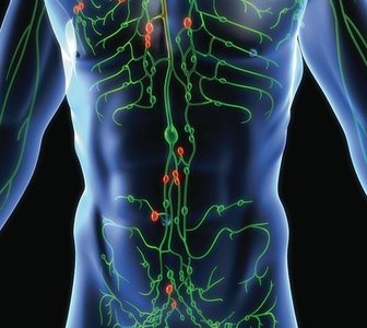
Lymph: Fluid in tissue spaces carrying proteins and other substances back to the blood.
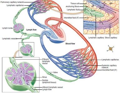
Lymphatic vessels: Permit one-way movement of lymph, beginning with microscopic, blind-ended lymphatic capillaries.
Lacteals: Specialized lymphatic capillaries in the intestinal wall that transport fats into the bloodstream.
Lymphatic Vessels and Ducts
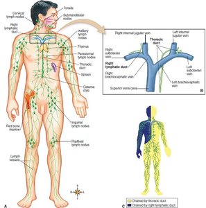
Right lymphatic duct: Drains lymph from the right upper extremity and right side of head, neck, and upper torso.
Thoracic duct: Largest lymphatic vessel; drains lymph from about three-fourths of the body. The cisterna chyli is an enlarged pouch along its course.
Lymphatic System in Fluid Homeostasis
The lymphatic system collects excess interstitial fluid and returns it to the bloodstream, preventing tissue swelling (edema) and maintaining blood volume.
Lymphatic Disorders
Lymphedema and Related Conditions
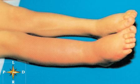
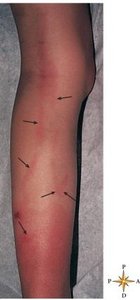
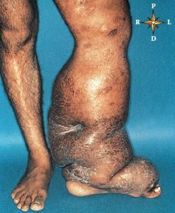
Lymphedema is swelling caused by blockage of lymphatic vessels. It can result from infection, surgery, or parasitic infestation.
Lymphangitis: Inflammation of lymphatic vessels, which may progress to septicemia.
Elephantiasis: Severe lymphedema of limbs due to parasite infestation.
Lymphoid Organs
Lymph Nodes
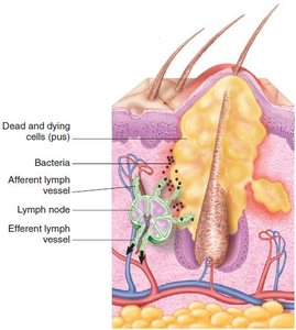
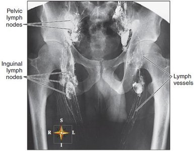
Lymph nodes are small, bean-shaped structures that filter lymph and are located in clusters along lymphatic vessels. They contain lymphoid tissue, which is rich in lymphocytes and other immune cells.
Functions: Defense (filtering pathogens) and white blood cell (WBC) formation.
Lymph flows into nodes via afferent vessels and exits via a single efferent vessel.
Lymphadenitis: Swelling and tenderness of lymph nodes, often due to infection.
Metastasis: Cancer cells can spread through lymphatic vessels to other body parts.
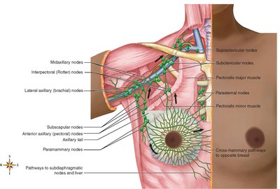
Lymphatic Drainage of the Breast
The breast contains an extensive network of lymphatic vessels and nodes, which is clinically significant in the spread of breast cancer and in post-surgical complications such as lymphedema.
Thymus
The thymus is a lymphoid organ located in the mediastinum. It is essential for the development of T lymphocytes (T cells) and the secretion of thymosins, hormones that regulate T-cell maturation. The thymus undergoes involution (replacement by fat) after childhood.
Tonsils
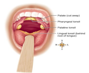
Tonsils are masses of lymphoid tissue located around the openings of the mouth and throat. They include the palatine, pharyngeal (adenoids), and lingual tonsils. Tonsils help protect against pathogens entering through the oral and nasal cavities but are prone to chronic infection and enlargement.
Spleen
The spleen is the largest lymphoid organ, located in the upper left abdomen. It filters blood, removes old red blood cells, and acts as a blood reservoir. Splenomegaly is enlargement of the spleen, often due to infection or disease.
Lymphoma
Lymphoma is a malignant tumor of lymph nodes. The two main types are Hodgkin disease and non-Hodgkin lymphoma, both of which cause painless lymph node enlargement and systemic symptoms.
Immunity: Innate and Adaptive Defenses
Function of the Immune System
The immune system protects the body from pathogens, foreign cells, and cancerous cells using a combination of nonspecific (innate) and specific (adaptive) mechanisms.
Innate (Nonspecific) Immunity
Innate immunity provides immediate, general defense against a wide range of pathogens. It includes physical barriers, phagocytic cells, and chemical mediators.
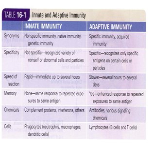
Innate Immunity | Adaptive Immunity |
|---|---|
Synonyms: Nonspecific, native, genetic immunity | Synonyms: Specific, acquired immunity |
Not specific—recognizes variety of nonself or abnormal cells and particles | Specific—recognizes only specific antigens on certain cells or particles |
Rapid—immediate up to several hours | Slower—several hours to several days |
No memory—same response to repeated exposures | Yes—enhanced response to repeated exposures |
Chemicals: Complement proteins, interferons, others | Chemicals: Antibodies, various signaling chemicals |
Cells: Phagocytes (neutrophils, macrophages, dendritic cells) | Cells: Lymphocytes (B cells and T cells) |
Mechanisms of Innate Defense
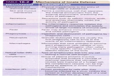
Mechanism | Description |
|---|---|
Mechanical and chemical barriers | Physical impediments to entry of foreign cells or substances |
Skin and mucous membranes | Continuous wall separating internal and external environments |
Secretions | Substances like sebum, mucus, acids, and enzymes inhibit pathogens |
Inflammation | Attracts immune cells, increases blood flow and vascular permeability |
Fever | Enhances immune reactions |
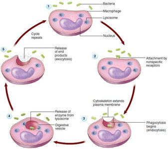
Phagocytosis | Ingestion and destruction of pathogens by cells |
Neutrophils | Short-lived phagocytes |
Macrophages | Long-lived phagocytes, often antigen-presenting cells |
Natural killer (NK) cells | Destroy any cell lacking a normal self-antigen |
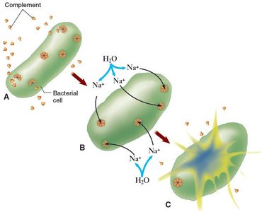
Complement | Protein cascade that produces cell lysis |
Interferon (IF) | Proteins released by virus-infected cells to inhibit viral replication |
Inflammatory Response
Inflammation is a key innate defense mechanism, characterized by heat, redness, pain, and swelling. It attracts immune cells, increases blood flow, and promotes healing.
Adaptive (Specific) Immunity
Adaptive immunity is the body's ability to recognize, respond to, and remember specific harmful substances. It involves B and T lymphocytes and provides long-lasting protection.
Inherited immunity: Present from birth.
Acquired immunity: Developed after exposure to antigens.
Types of Acquired Immunity
Type | How Acquired | Example |
|---|---|---|
Natural Active | Exposure to disease | Recovery from infection |
Natural Passive | Mother to child (placenta, milk) | Maternal antibodies |
Artificial Active | Vaccination | Immunization shots |
Artificial Passive | Injection of antibodies | Antiserum administration |
Immune System Molecules
Cytokines: Signaling proteins that regulate immune responses.
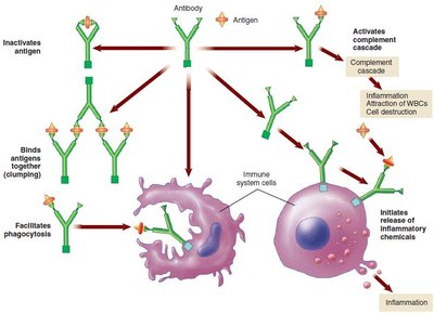
Antibodies: Proteins that bind specific antigens, forming antigen-antibody complexes. Functions include neutralizing toxins, agglutinating cells, and promoting phagocytosis.
Complement Proteins and Cascade
Complement proteins are a group of plasma proteins that, when activated, enhance immune responses by lysing foreign cells and promoting inflammation.
Activation occurs when antibodies bind antigens, exposing complement-binding sites.
The complement cascade results in the formation of protein rings that create holes in foreign cell membranes, causing cell lysis.
Immune System Cells
Phagocytes
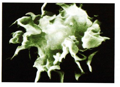
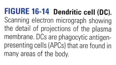
Phagocytes ingest and destroy foreign substances. Major types include neutrophils, monocytes (which become macrophages), and dendritic cells (DCs). Macrophages and DCs also act as antigen-presenting cells (APCs).
Lymphocytes
Lymphocytes are the most numerous immune cells and include natural killer (NK) cells, B cells, and T cells. NK cells are part of innate immunity, while B and T cells are central to adaptive immunity.
Natural Killer (NK) Cells: Destroy virus-infected and tumor cells without prior exposure.
B Cells: Develop in bone marrow, produce antibodies (humoral immunity), and form plasma and memory cells.
T Cells: Develop in the thymus, mediate cell-mediated immunity, and differentiate into cytotoxic, helper, and regulatory T cells.
Immune System Disorders
Hypersensitivity
Hypersensitivity is an excessive or inappropriate immune response, such as allergies (immediate or delayed) and contact dermatitis.
Autoimmunity
Autoimmunity is an immune response against self-antigens, leading to diseases like systemic lupus erythematosus (SLE).
Alloimmunity
Alloimmunity (isoimmunity) is an excessive reaction to antigens from another individual, important in pregnancy and tissue transplants.
Immune Deficiency
Congenital immune deficiency: Present at birth, e.g., severe combined immune deficiency (SCID).
Acquired immune deficiency: Develops after birth, e.g., AIDS caused by HIV infection of T cells.
Review Questions
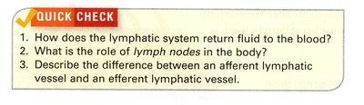
What is the role of lymph nodes in the body?
Describe the difference between afferent and efferent lymphatic vessels.
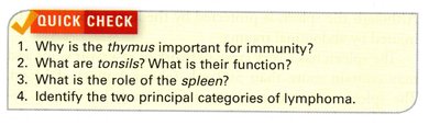
Why is the thymus important for immunity?
What are the two principal categories of lymphoma?
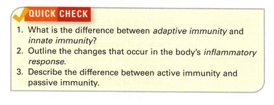
What is the difference between adaptive and innate immunity?
Describe the difference between active and passive immunity.
