 Back
BackLymphatic System and Immunity: Structure, Function, and Clinical Significance
Study Guide - Smart Notes
 Tailored notes based on your materials, expanded with key definitions, examples, and context.
Lymphatic System: Structure and Function
Overview of the Lymphatic System
The lymphatic system is a vital component of the human body, responsible for maintaining fluid balance, filtering pathogens, and supporting immune function. It consists of a network of vessels, nodes, and organs that transport lymph—a fluid containing proteins, waste products, and immune cells—back to the bloodstream.
Main Functions: Fluid homeostasis, immune defense, absorption of fats from the digestive tract.
Main Structures: Lymphatic vessels, lymph nodes, thymus, tonsils, spleen.
Lymph: Specialized fluid formed from interstitial fluid, carrying proteins and other substances back to the blood.
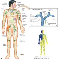
Lymphatic Vessels
Lymphatic vessels permit only one-way movement of lymph, ensuring that excess fluid and proteins are returned to the circulatory system. Lymphatic capillaries are tiny, blind-ended tubes distributed throughout tissue spaces, composed of a single layer of simple squamous epithelium.
Lacteals: Specialized lymphatic capillaries in the intestinal wall that transport fats.
Right Lymphatic Duct: Drains lymph from the right upper extremity and right side of head, neck, and upper torso.
Thoracic Duct: Largest lymphatic vessel, drains lymph from about three-fourths of the body; contains the cisterna chyli, a storage area for lymph.
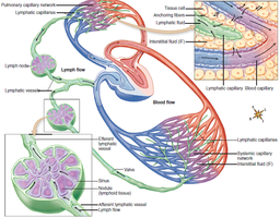
Fluid Homeostasis and Lymph Formation
Fluid filtered from blood plasma that is not reabsorbed by blood vessels drains into lymphatic vessels. This process prevents the accumulation of excess tissue fluid and maintains homeostasis.
Interstitial Fluid: Fluid in tissue spaces before it enters lymphatic vessels.
Lymph Nodes: Filter lymphatic fluid before it is returned to the bloodstream.
Clinical Conditions of the Lymphatic System
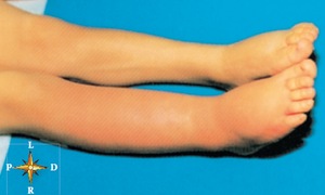
Lymphedema
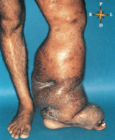
Lymphedema is swelling of tissues caused by blockage of lymphatic vessels, leading to accumulation of lymph. It can result from infection, surgery, or parasitic infestation.
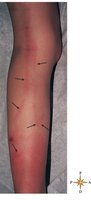
Lymphangitis: Inflammation of lymphatic vessels, may progress to septicemia.
Elephantiasis: Severe lymphedema caused by parasite infestation (Filaria worms).
Lymphoid Organs and Their Functions
Lymph Nodes
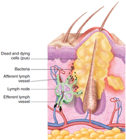
Lymph nodes are clusters of lymphoid tissue located along lymphatic vessels. They filter lymph, provide immune defense, and support white blood cell formation.
Flow of Lymph: Enters node via afferent vessels, exits via a single efferent vessel.
Lymphadenitis: Swelling and tenderness of lymph nodes.
Metastasis: Cancer cells can spread through lymphatic vessels.
Lymphangiogram
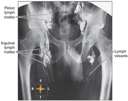
A lymphangiogram is a radiographic procedure using dye to visualize lymphatic vessels and nodes, often used to assess lymphatic drainage and detect blockages or tumors.
Lymphatic Drainage of the Breast
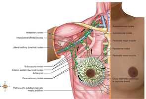
The breast is drained by a network of lymphatic vessels and nodes, which are important in the spread and treatment of breast cancer.
Tonsils
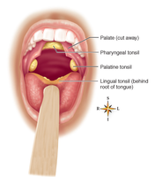
Tonsils are masses of lymphoid tissue located around the openings of the mouth and throat. They serve as the first line of defense against pathogens entering through the oral and nasal cavities.
Palatine Tonsils: Located on each side of the throat.
Pharyngeal Tonsils (Adenoids): Near the posterior opening of the nasal cavity.
Lingual Tonsils: Near the base of the tongue.
Spleen
The spleen is the largest lymphoid organ, located in the upper left quadrant of the abdomen. It functions in phagocytosis of bacteria and old red blood cells, acts as a blood reservoir, and stores iron salvaged from hemoglobin.
Splenectomy: Surgical removal of the spleen, often due to trauma.
Splenomegaly: Enlargement of the spleen.
Immune System: Mechanisms and Cells
Function of the Immune System
The immune system protects the body from pathogens, foreign tissue cells, and cancerous cells. It consists of defensive cells and molecules, rather than organs.
Nonspecific Immunity (Innate): Rapid response to a variety of nonself cells and particles.
Specific Immunity (Adaptive): Ability to recognize, respond to, and remember harmful substances.
Innate Immunity
Innate immunity includes mechanical barriers (skin), chemical barriers (tears, mucus), and cellular responses (inflammation, phagocytosis).
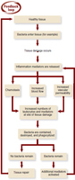
Inflammation: Attracts immune cells, increases blood flow and vascular permeability, promotes movement of WBCs to site of injury.
Signs: Heat, redness, pain, swelling.
Adaptive Immunity
Adaptive immunity is specific and involves memory. It can be inherited or acquired, and is further classified as active or passive, natural or artificial.
Active Immunity: Develops after exposure to antigen (infection or vaccination).
Passive Immunity: Acquired from another individual (maternal antibodies, immunotherapy).
Immune System Molecules
Key molecules include cytokines and antibodies. Antibodies are protein compounds with specific combining sites that attach to antigens, forming antigen-antibody complexes.
Functions: Neutralize toxins, agglutinate enemy cells, promote phagocytosis.
Cytokines: Interleukins, used by WBCs for communication.
Complement Proteins
Complement proteins are present in blood in an inactive state. When activated, they trigger a cascade of reactions that create holes in the plasma membrane of foreign cells, leading to cell lysis.
Roles: Attract immune cells, activate immune cells, mark foreign cells for destruction, increase blood vessel permeability.
Immune System Cells
The immune system is composed of phagocytes and lymphocytes, each with specialized functions.
Phagocytes: Neutrophils, monocytes (macrophages), dendritic cells; ingest and destroy foreign cells.
Lymphocytes: B cells, T cells, natural killer cells.
B Cell Development and Function
B cells develop in the bone marrow and migrate to lymph nodes. Upon activation by antigen, they differentiate into plasma cells (which secrete antibodies) and memory cells (which provide long-term immunity).
Humoral Immunity: Indirectly produced by B cells via circulating antibodies.
T Cell Development and Function
T cells develop in the thymus and migrate to lymph nodes. Upon activation, they differentiate into effector cells (cytotoxic, helper, regulatory) and memory cells.
Cell-Mediated Immunity: Produced by T cells through direct action on infected or tumor cells.
Immune System Disorders
Hypersensitivity
Hypersensitivity is an inappropriate or excessive immune response, including allergies (response to harmless antigens), autoimmunity (response to self-antigens), and alloimmunity (response to antigens from another human).
Immediate Allergic Responses: Usually involve humoral immunity.
Delayed Allergic Responses: Usually involve cell-mediated immunity.
Autoimmunity
Autoimmunity results from excessive response to self-antigens, causing diseases such as systemic lupus erythematosus (SLE).
Immune System Deficiency
Immune deficiencies may be congenital (improper lymphocyte development) or acquired (AIDS, caused by HIV infection of T cells).
SCID: Severe combined immune deficiency, caused by disruption of stem cell development.
AIDS: Acquired immunodeficiency syndrome, treated with antiviral drugs.
Type of Immunity | Source | Duration | Example |
|---|---|---|---|
Natural Active | Exposure to pathogen | Long-term | Measles infection |
Natural Passive | Maternal antibodies | Short-term (3-6 months) | Placental transfer |
Artificial Active | Vaccination | Long-term | Immunization |
Artificial Passive | Immunotherapy | Short-term | Gamma globulin injection |
Additional info: The notes above expand on the original content by providing definitions, examples, and clinical context for each topic, ensuring completeness and academic quality for college-level study.
