 Back
BackLymphatic System and Lymphoid Organs: Structure, Function, and Clinical Aspects
Study Guide - Smart Notes
 Tailored notes based on your materials, expanded with key definitions, examples, and context.
Lymphatic System Overview
General Functions and Components
The lymphatic system is essential for returning fluids leaked from blood vessels back to the blood and for providing the structural basis of the immune system. It consists of a network of lymphatic vessels, lymph (the fluid within these vessels), and lymph nodes that cleanse the lymph. Lymphoid organs and tissues house phagocytic cells and lymphocytes, supporting immune function.
Lymphatic vessels: Drainage network for interstitial fluid.
Lymph: Fluid that enters lymphatic vessels.
Lymph nodes: Filter lymph and house immune cells.
Lymphoid organs: Include spleen, thymus, tonsils, lymph nodes, and other tissues.
Structural Relationship of Lymphatic and Blood Capillaries
Lymphatic capillaries are closely associated with blood capillaries, forming a structural relationship that facilitates fluid exchange between tissues and the vascular system.
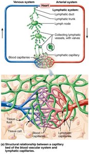
Distribution and Structure of Lymphatic Vessels
Lymphatic Capillaries
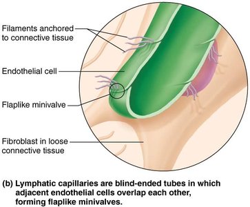
Lymphatic capillaries are blind-ended vessels that weave between tissue cells and blood capillaries. They are absent from bones, teeth, and bone marrow, but present in limited locations in the CNS. Their permeability allows uptake of larger molecules and particles, such as proteins, cell debris, pathogens, and cancer cells.
Minivalves: Formed by overlapping endothelial cells, allowing one-way entry of fluid.
Anchoring filaments: Attach minivalves to connective tissue, opening them with increased ECF volume.
Lacteals: Specialized capillaries in intestinal mucosa that absorb digested fat (chyle).
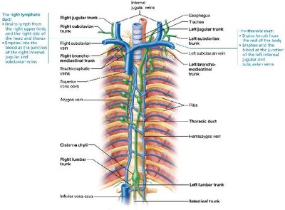
Larger Lymphatic Vessels
Lymphatic capillaries drain into larger collecting vessels, trunks, and ducts. These vessels have thinner walls and more internal valves than veins, and they anastomose more frequently. Collecting vessels in skin travel with superficial veins, while deep vessels travel with arteries.
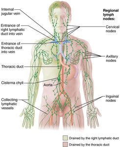
Lymphatic trunks: Drain large regions of the body (lumbar, bronchomediastinal, subclavian, jugular, intestinal).
Lymphatic ducts: Right lymphatic duct drains right upper arm and right side of head/thorax; thoracic duct drains the rest of the body.
Cisterna chyli: Enlarged sac at the base of the thoracic duct in about half of individuals.
Lymph Transport and Clinical Aspects
Lymph Transport Mechanisms
The lymphatic system is a low-pressure system, similar to veins. Lymph is propelled by skeletal muscle contraction, thoracic pressure changes during breathing, valves preventing backflow, arterial pulsations, and smooth muscle contractions in vessel walls. Physical activity increases lymph flow, while immobilization slows it, aiding healing.
Clinical Imbalances
Lymphangitis: Painful red lines under the skin due to inflamed lymphatic vessels.
Lymphedema: Severe localized edema caused by blockage or removal of lymphatic vessels.
Lymphoid Cells, Tissues, and Organs
Lymphoid Cells
Lymphoid cells include immune system cells (lymphocytes, macrophages, dendritic cells) and supporting cells (reticular cells).
Lymphocytes: T cells manage immune response and attack infected cells; B cells produce plasma cells that secrete antibodies.
Macrophages: Phagocytize foreign substances and activate T cells.
Dendritic cells: Capture antigens and deliver them to lymph nodes.
Reticular cells: Produce reticular fibers (stroma) for structural support.
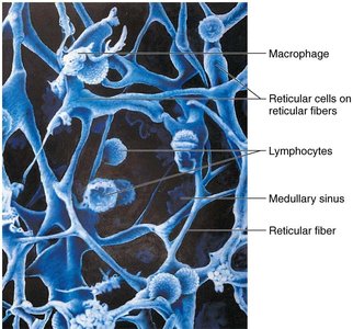
Lymphoid Tissue
Lymphoid tissue houses and provides proliferation sites for lymphocytes, offering surveillance vantage points. It is largely composed of reticular connective tissue, with macrophages living on reticular fibers and spaces for lymphocytes.
Types of Lymphoid Tissue
Diffuse lymphoid tissue: Loose arrangement found in most organs.
Lymphoid follicles (nodules): Spherical bodies with germinal centers of proliferating B cells; found in nodes, Peyer's patches, and appendix.
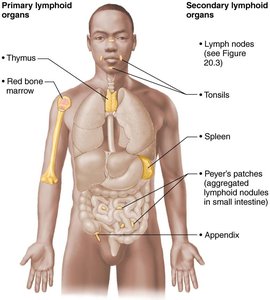
Primary and Secondary Lymphoid Organs
Primary: Sites of T and B cell maturation (red bone marrow, thymus).
Secondary: Sites where mature lymphocytes encounter antigens (nodes, spleen, MALT, diffuse tissues).
Lymph Nodes
Structure and Function
Lymph nodes are principal secondary lymphoid organs, found in clusters along lymphatic vessels. They filter lymph and provide sites for immune activation.
Cleansing: Macrophages remove microorganisms and debris.
Immune activation: Lymphocytes mount attacks against antigens.
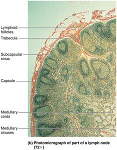
Internal Structure
Lymph nodes are bean-shaped, less than 2.5 cm, with an external capsule and internal trabeculae dividing compartments. Two regions: cortex (with follicles and germinal centers) and medulla (with medullary cords and sinuses).
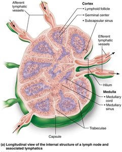
Lymph Circulation
Lymph enters via afferent vessels, passes through sinuses, and exits via efferent vessels at the hilum.
Fewer efferent vessels cause stagnation, allowing immune cells time to act.

Clinical Imbalances
Buboes: Swollen, tender lymph nodes overwhelmed by pathogens (e.g., bubonic plague).
Cancer metastasis: Cancer cells trapped in nodes, causing painless swelling.
Spleen
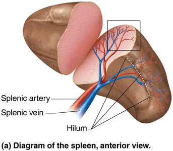
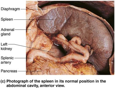
Structure and Functions
The spleen is the largest lymphoid organ, located in the left abdominal cavity. It is served by the splenic artery and vein at the hilum. Functions include lymphocyte proliferation, immune surveillance, and cleansing blood of aged cells and platelets.
Stores breakdown products of RBCs (e.g., iron).
Stores platelets and monocytes.
Site of fetal erythrocyte production.
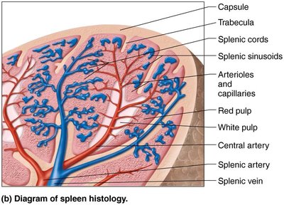
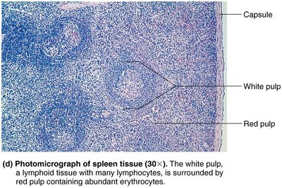
Histological Components
White pulp: Site of immune function, clusters around central arteries.
Red pulp: Site of destruction of old blood cells and pathogens, rich in RBCs and macrophages.
Clinical Imbalances
Splenic rupture: May require splenectomy; liver and bone marrow compensate for lost functions.
Regeneration: In children, spleen can regenerate if a portion remains.
MALT (Mucosa-Associated Lymphoid Tissue)
Structure and Function
MALT consists of lymphoid tissues in mucous membranes throughout the body, protecting against pathogens entering via respiratory, genitourinary, and digestive tracts. Largest collections are found in tonsils, Peyer's patches, and appendix.
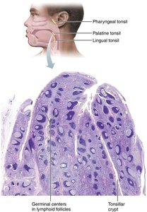
Tonsils
Form a ring of lymphatic tissue around the pharynx.
Named by location: palatine, lingual, pharyngeal (adenoids), tubal.
Function: Gather and remove pathogens in food or air.
Contain follicles with germinal centers and tonsillar crypts for trapping bacteria.
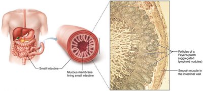
Peyer's Patches
Clusters of lymphoid follicles in the distal small intestine.
Destroy bacteria and generate memory lymphocytes.
Appendix
Offshoot of the large intestine, contains many lymphoid follicles.
Functions similar to Peyer's patches: destroy bacteria and generate memory lymphocytes.
Thymus
Structure and Function
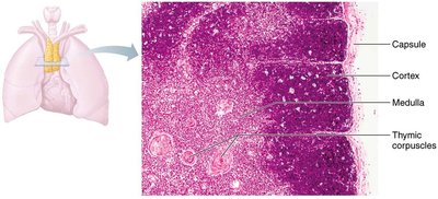
The thymus is a bilobed lymphoid organ in the inferior neck, extending into the mediastinum. It is the site of T cell maturation, most active in childhood, and gradually atrophies with age. It produces immunocompetent cells throughout life.
Broken into lobules with cortex (rapidly dividing lymphocytes) and medulla (fewer lymphocytes, thymic corpuscles).
Regulatory T cells develop in thymic corpuscles, preventing autoimmunity.
No follicles (lacks B cells), does not directly fight antigens, stroma made of epithelial cells.
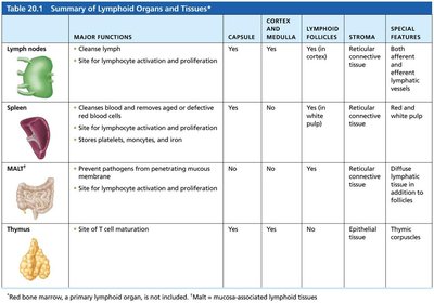
Summary Table: Lymphoid Organs and Tissues
The following table summarizes the major functions, structural features, and special characteristics of lymphoid organs and tissues:
Organ/Tissue | Major Functions | Capsule | Cortex & Medulla | Lymphoid Follicles | Stroma | Special Features |
|---|---|---|---|---|---|---|
Lymph nodes | Cleanses lymph; site for lymphocyte activation and proliferation | Yes | Yes | Yes (in cortex) | Reticular connective tissue | Both afferent and efferent lymphatic vessels |
Spleen | Cleanses blood; removes aged/defective blood cells; site for lymphocyte activation; stores platelets, monocytes, iron | Yes | Yes | Yes (in white pulp) | Reticular connective tissue | White and red pulp |
MALT* | Prevents pathogens from penetrating mucosa; site for lymphocyte activation and proliferation | No | No | Yes | Diffuse reticular connective tissue | Collections in mucosa |
Thymus | Site of T cell maturation | Yes | Yes | No | Epithelial tissue | Thymic corpuscles |
Developmental Aspects of the Lymphatic System
Embryonic Development
Lymphatic vessels and main clusters of lymph nodes appear by week 5 of embryonic development, arising as lymph sacs from developing veins. Jugular lymph sacs form the right lymphatic duct and thoracic duct. Lymphatic organs (except thymus) arise from mesodermal mesenchymal cells, while the thymus forms from endodermal origin as an outgrowth of the pharynx.
Except for spleen and tonsils, lymphoid organs are poorly developed at birth.
High numbers of lymphocytes appear after birth, paralleling immune system maturation.
