 Back
BackLymphatic System: Structure, Function, and Lymphoid Organs
Study Guide - Smart Notes
 Tailored notes based on your materials, expanded with key definitions, examples, and context.
Lymphatic System Overview
Introduction to the Lymphatic System
The lymphatic system is a vital component of the circulatory and immune systems. It returns fluids that have leaked from blood vessels back to the bloodstream and provides the structural basis for immune defense.
Key Functions: Maintains blood volume, removes foreign matter, and mobilizes the immune system.
Main Components: Network of lymphatic vessels, lymph (fluid), and lymph nodes.
Lymphatic System Functions
Fluid Recovery: About 3 liters of fluid per day remain in tissues after capillary exchange; lymphatic vessels return this interstitial fluid and leaked plasma proteins to the blood.
Immune Defense: Lymphoid organs house phagocytic cells and lymphocytes, which defend against pathogens.
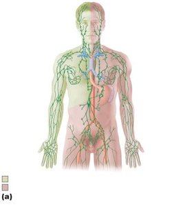
Lymphatic Vessels: Structure and Distribution
Types of Lymphatic Vessels
Lymphatic vessels form a one-way system that transports lymph toward the heart.
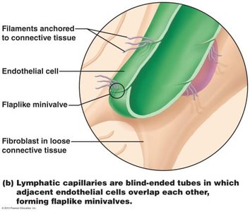
Lymphatic Capillaries: Highly permeable, take up proteins, cell debris, pathogens, and cancer cells. Endothelial cells overlap to form one-way minivalves, opened by increased tissue pressure.
Collecting Lymphatic Vessels: Similar to veins but with thinner walls and more internal valves. They anastomose more frequently and are supplied by vasa vasorum.
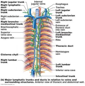
Lymphatic Trunks and Ducts: Trunks drain large regions and are named for the areas they serve (e.g., lumbar, bronchomediastinal, subclavian, jugular, intestinal). Ducts include the right lymphatic duct and thoracic duct.
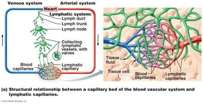
Lymphatic Capillaries and Lacteals
Absent From: Bones, teeth, bone marrow, and CNS.
Lacteals: Specialized capillaries in intestinal mucosa that absorb digested fat and deliver chyle to the blood.
Lymphatic Ducts
Right Lymphatic Duct: Drains right upper arm and right side of head and thorax.
Thoracic Duct: Drains the rest of the body; empties into venous circulation at the junction of internal jugular and subclavian veins.
Lymph Transport Mechanisms
Lymph is propelled by several mechanisms:
Milking action of skeletal muscle
Pressure changes during breathing
Valves to prevent backflow
Pulsations of nearby arteries
Contractions of smooth muscle in vessel walls
Lymphoid Cells and Tissues
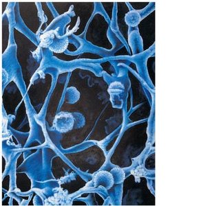
Lymphoid Cells
Lymphoid cells are essential for immune defense.
Lymphocytes: Main warriors of the immune system, arising in red bone marrow. Two main types:
T cells (T lymphocytes): Manage immune response, attack and destroy infected cells.
B cells (B lymphocytes): Produce plasma cells that secrete antibodies, marking antigens for destruction.
Macrophages: Phagocytize foreign substances and help activate T cells.
Dendritic Cells: Capture antigens and deliver them to lymph nodes, activating T cells.
Reticular Cells: Produce reticular fiber stroma that supports other cells in lymphoid organs.
Lymphoid Tissue
Lymphoid tissue provides a site for lymphocyte proliferation and immune surveillance.
Diffuse Lymphoid Tissue: Loose arrangement of cells and fibers, found in nearly every body organ.
Lymphoid Follicles (Nodules): Solid, spherical bodies of tightly packed lymphoid cells and reticular fibers, with germinal centers of proliferating B cells.
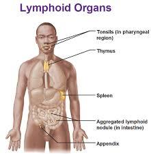
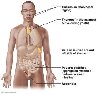
Lymphoid Organs
Major Lymphoid Organs
Lymphoid organs provide structural support and house immune cells.
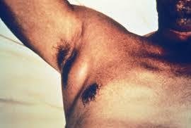
Lymph Nodes: Principal lymphoid organs, embedded in connective tissue and clustered along lymphatic vessels. Located near body surface in inguinal, axillary, and cervical regions.
Spleen: Largest lymphoid organ, site of lymphocyte proliferation, immune surveillance, and blood cleansing.
Thymus: Site of T lymphocyte maturation, most active during childhood.
Tonsils: Simplest lymphoid organs, form a ring of lymphatic tissue around the pharynx.
Peyer's Patches and Appendix: Aggregated lymphoid nodules in the intestine, destroy bacteria and generate memory lymphocytes.

Lymph Nodes: Structure and Function
Lymph nodes filter lymph and activate the immune system.
Structure: Bean-shaped, external fibrous capsule, trabeculae divide node into compartments. Two regions: cortex (follicles with B cells, deep cortex with T cells) and medulla (cords with B cells, T cells, plasma cells; sinuses with macrophages).
Function:
Filter lymph—macrophages destroy microorganisms and debris.
Immune activation—lymphocytes mount attack against antigens.
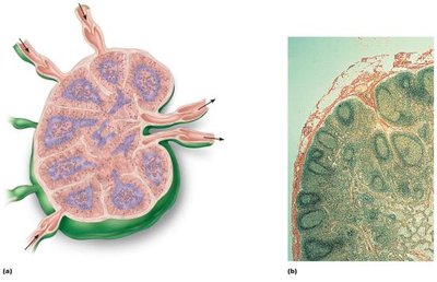
Lymph Node Circulation
Lymph enters via afferent vessels, travels through sinuses, and exits at the hilum via efferent vessels.
Fewer efferent vessels cause stagnation, allowing time for immune cells to function.
Lymph Node Imbalances
Inflamed lymph nodes (swollen glands or buboes) indicate infection.
Lymph nodes can become secondary cancer sites.
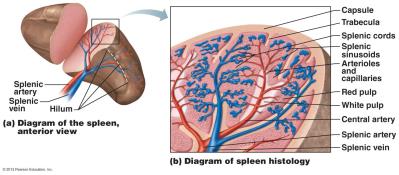
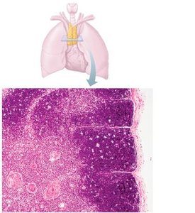
Spleen
Structure and Functions of the Spleen
The spleen is the largest lymphoid organ, located in the left upper abdomen.
Functions:
Site of lymphocyte proliferation and immune response
Cleanses blood of aged cells and platelets
Stores breakdown products of RBCs (e.g., iron)
Stores blood platelets and monocytes
May produce erythrocytes in fetus
Structure: Two areas:
White pulp: Lymphocytes involved in immune functions
Red pulp: Rich in RBCs and macrophages for disposal of worn-out cells and pathogens
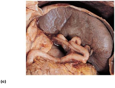
Spleen Imbalances
The spleen can rupture easily but may repair itself. If removed, liver and bone marrow compensate. In children, the spleen can regenerate if a portion remains.
Thymus
Structure and Function of the Thymus
The thymus is essential for T lymphocyte maturation and is most active during childhood.
Location: Inferior neck, partially overlies the heart.
Structure: Lacks follicles (no B cells), functions strictly in T cell maturation, isolated by blood-thymus barrier.
Development: Increases in size during childhood, atrophies after adolescence but continues to produce immunocompetent cells.
Mucosa-Associated Lymphoid Tissue (MALT)
Definition and Locations
MALT consists of lymphoid tissues in mucous membranes throughout the body, protecting against pathogens.
Major Collections: Tonsils, Peyer's patches, appendix, and tissues in respiratory, genitourinary, and digestive tracts.
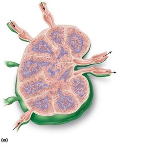
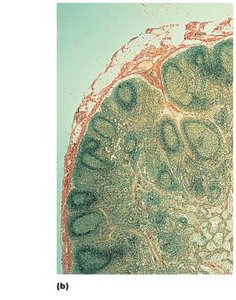
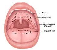
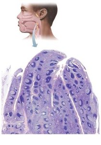
Tonsils
Structure: Form a ring of lymphatic tissue around the pharynx. Types include palatine, lingual, pharyngeal (adenoids), and tubal tonsils.
Function: Gather and remove pathogens in food or air. Contain follicles with germinal centers and tonsillar crypts that trap and destroy bacteria.
Peyer's Patches and Appendix
Peyer's Patches: Clusters of lymphoid follicles in the wall of the distal small intestine.
Appendix: Contains similar lymphoid structures.
Function: Destroy bacteria and generate memory lymphocytes.
Developmental Aspects of the Lymphatic System
Embryonic Development
Lymphatic vessels and main clusters of lymph nodes begin forming by the 5th week of embryonic development, arising as lymph sacs from developing veins.
Lymphatic organs arise from mesoderm, except the thymus (endoderm).
Postnatal Development
Except for spleen and tonsils, lymphoid organs are poorly developed at birth.
High numbers of lymphocytes after birth; their development parallels maturation of the immune system.
