 Back
BackLymphatic System Study Guide – Anatomy & Physiology
Study Guide - Smart Notes
 Tailored notes based on your materials, expanded with key definitions, examples, and context.
Q1. Label the lymphatic structures on the image below.
Background
Topic: Lymphatic System Anatomy
This question tests your knowledge of the major lymphatic structures in the human body, including lymphatic vessels, lymph nodes, and associated organs.
Key Terms:
Lymphatic vessels
Lymph nodes
Thymus
Spleen
Tonsils
Thoracic duct
Right lymphatic duct
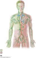
Step-by-Step Guidance
Observe the image and identify the green-colored vessels and nodes, which represent the lymphatic system.
Locate the major clusters of lymph nodes (cervical, axillary, and inguinal regions).
Identify the thoracic duct and right lymphatic duct, which are the main lymphatic drainage vessels.
Find the spleen, thymus, and tonsils, which are key lymphatic organs.
Label each structure based on its anatomical position and appearance in the diagram.
Try solving on your own before revealing the answer!
Final Answer:
Lymphatic vessels: Green lines throughout the body
Lymph nodes: Small green oval structures clustered in the neck (cervical), armpit (axillary), and groin (inguinal) regions
Thoracic duct: Large green vessel running along the left side of the vertebral column
Right lymphatic duct: Small green vessel draining the right upper quadrant
Spleen: Green organ on the left side of the abdomen
Thymus: Green organ in the upper chest, above the heart
Tonsils: Green structures in the throat region
These are the main lymphatic structures visible in the diagram.
Q2. Label the lymphatic structures on the image below.
Background
Topic: Lymphatic Vessels of the Thorax
This question focuses on the lymphatic drainage pathways in the thoracic region, including the thoracic duct and its relationship to major veins.
Key Terms:
Thoracic duct
Right lymphatic duct
Subclavian vein
Brachiocephalic vein
Lymphatic trunks
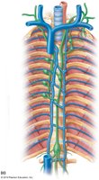
Step-by-Step Guidance
Examine the image for the large blue and green vessels in the thoracic cavity.
Identify the thoracic duct, which is the main lymphatic vessel running superiorly in the thorax.
Locate the right lymphatic duct, which is much shorter and drains the right upper quadrant.
Find the subclavian and brachiocephalic veins, which are closely associated with the lymphatic ducts.
Label the lymphatic trunks that feed into these ducts.
Try solving on your own before revealing the answer!
Final Answer:
Thoracic duct: Large green vessel ascending in the thorax
Right lymphatic duct: Small green vessel on the right side
Subclavian vein: Large blue vein under the clavicle
Brachiocephalic vein: Blue vein formed by the union of subclavian and internal jugular veins
Lymphatic trunks: Green vessels feeding into the ducts
These are the main lymphatic structures in the thoracic region.
Q3. Label the lymphatic structures on the image below.
Background
Topic: Lymphatic Organs
This question tests your ability to identify the primary and secondary lymphatic organs, such as the thymus, spleen, and tonsils.
Key Terms:
Thymus
Spleen
Tonsils
Lymph nodes
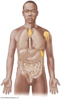
Step-by-Step Guidance
Look for the highlighted yellow structures in the image, which represent lymphatic organs.
Identify the thymus in the upper chest, just above the heart.
Locate the spleen on the left side of the abdomen, near the stomach.
Find the tonsils in the throat region and any visible lymph nodes.
Label each organ based on its anatomical location.
Try solving on your own before revealing the answer!
Final Answer:
Thymus: Yellow organ in the upper chest
Spleen: Yellow organ on the left side of the abdomen
Tonsils: Yellow structures in the throat
Lymph nodes: Small yellow clusters in the axillary and inguinal regions
These are the main lymphatic organs shown in the diagram.
Q4. Label the lymph node structures below.
Background
Topic: Lymph Node Microanatomy
This question tests your understanding of the internal structure of a lymph node, including its regions and pathways for lymph flow.
Key Terms:
Capsule
Cortex
Medulla
Lymphoid follicle
Germinal center
Trabeculae
Afferent and efferent lymphatic vessels
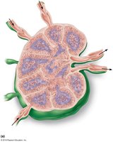
Step-by-Step Guidance
Identify the outermost layer (capsule) surrounding the lymph node.
Locate the cortex, which is the outer region beneath the capsule, containing lymphoid follicles.
Find the medulla, the inner region with medullary cords and sinuses.
Spot the lymphoid follicles and their germinal centers within the cortex.
Label the trabeculae, which are extensions of the capsule into the node, and the afferent/efferent vessels.
Try solving on your own before revealing the answer!
Final Answer:
Capsule: Outermost layer
Cortex: Outer region beneath capsule
Medulla: Inner region
Lymphoid follicle: Spherical structures in cortex
Germinal center: Light area within follicle
Trabeculae: Extensions of capsule
Afferent vessels: Entering the node
Efferent vessels: Exiting the node
These are the main structures of a lymph node.
Q5. Label the following structure on the image below: antigen binding site, antigen, antigenic determinant, heavy chain (variable and constant), light chain (variable and constant), hinge region, stem region, disulfide bridge.
Background
Topic: Antibody Structure
This question tests your understanding of the molecular structure of antibodies (immunoglobulins) and the regions involved in antigen recognition and immune function.
Key Terms:
Antigen binding site
Antigen
Antigenic determinant (epitope)
Heavy chain (variable and constant regions)
Light chain (variable and constant regions)
Hinge region
Stem region (Fc region)
Disulfide bridge
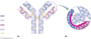
Step-by-Step Guidance
Identify the Y-shaped antibody molecule in the image.
Locate the tips of the Y, which are the antigen binding sites.
Find the antigen and its antigenic determinant (epitope) interacting with the binding site.
Distinguish between the heavy and light chains, and their variable and constant regions.
Label the hinge region, stem region, and disulfide bridges connecting the chains.
Try solving on your own before revealing the answer!
Final Answer:
Antigen binding site: Tip of each arm of the Y
Antigen: Structure binding to the tip
Antigenic determinant: Specific region on the antigen
Heavy chain: Longer polypeptide, with variable (top) and constant (bottom) regions
Light chain: Shorter polypeptide, with variable (top) and constant (bottom) regions
Hinge region: Flexible area at the base of the Y arms
Stem region: Bottom of the Y (Fc region)
Disulfide bridge: Yellow bonds connecting chains
These are the main structural features of an antibody molecule.
Q6. Fill out the table below.
Background
Topic: Histology of Lymphatic Organs
This question asks you to identify and describe the microscopic structures of the lymph node, spleen, and tonsil.
Key Terms:
Lymph node: Capsule, cortex, medulla, lymphoid follicle, germinal center, trabeculae
Spleen: White pulp, red pulp
Tonsil: Germinal center, crypt
Step-by-Step Guidance
For each organ, list the structures you need to label or draw (as given in the table).
Recall the location and function of each structure (e.g., cortex is the outer region of a lymph node).
Sketch or label the structures based on their typical histological appearance.
For the spleen, distinguish between white pulp (lymphatic tissue) and red pulp (blood-filled sinuses).
For the tonsil, identify the germinal center and crypts (invaginations).
Try solving on your own before revealing the answer!
Final Answer:
Lymph Node: Capsule (outer layer), cortex (outer region), medulla (inner region), lymphoid follicle (in cortex), germinal center (within follicle), trabeculae (extensions of capsule)
Spleen: White pulp (lymphatic tissue around central arteries), red pulp (blood-filled sinuses)
Tonsil: Germinal center (within lymphoid follicle), crypt (invagination of surface epithelium)
These are the main histological features to label or draw for each organ.
Q7. What is lymph?
Background
Topic: Lymphatic Fluid
This question tests your understanding of the composition and function of lymph in the body.
Key Terms:
Lymph: Interstitial fluid collected by lymphatic vessels
Plasma, interstitial fluid
Step-by-Step Guidance
Recall that lymph originates as interstitial fluid that enters lymphatic capillaries.
Consider the components of lymph (water, proteins, white blood cells, etc.).
Think about the role of lymph in returning fluid to the bloodstream and in immune surveillance.
Try solving on your own before revealing the answer!
Final Answer:
Lymph is a clear fluid that originates as interstitial fluid, which is collected from tissues by lymphatic capillaries. It contains water, proteins, white blood cells, and sometimes fats, and plays a key role in returning fluid to the bloodstream and in immune defense.
Q8. What factors are involved in the flow of lymphatic fluid?
Background
Topic: Lymph Flow Physiology
This question tests your knowledge of the mechanisms that propel lymph through the lymphatic system.
Key Terms:
Skeletal muscle pump
Respiratory pump
Valves in lymphatic vessels
Pressure gradients
Step-by-Step Guidance
Recall that lymphatic vessels lack a central pump like the heart.
Think about how skeletal muscle contractions help move lymph.
Consider the role of breathing (respiratory pump) in lymph flow.
Remember that valves prevent backflow in lymphatic vessels.
Try solving on your own before revealing the answer!
Final Answer:
Skeletal muscle contractions
Respiratory movements (pressure changes during breathing)
Valves in lymphatic vessels
Arterial pulsations and smooth muscle contractions in vessel walls
These factors work together to move lymph toward the venous system.
Q9. What name is given to the terminal duct draining most of the body?
Background
Topic: Lymphatic Ducts
This question tests your knowledge of the major lymphatic drainage pathways.
Key Terms:
Thoracic duct
Right lymphatic duct
Step-by-Step Guidance
Recall the two main lymphatic ducts: thoracic duct and right lymphatic duct.
Determine which duct drains the majority of the body (left side, both lower limbs, abdomen, left thorax, left arm, left side of head and neck).
Remember the anatomical location and drainage area of each duct.
Try solving on your own before revealing the answer!
Final Answer:
The thoracic duct is the terminal duct that drains lymph from most of the body into the left subclavian vein.
Q10. What is the cisterna chyli?
Background
Topic: Lymphatic Anatomy
This question tests your understanding of the structure and function of the cisterna chyli in the lymphatic system.
Key Terms:
Cisterna chyli
Thoracic duct
Lymph from lower limbs and abdomen
Step-by-Step Guidance
Recall the location of the cisterna chyli (inferior end of the thoracic duct, in the abdomen).
Think about its function as a collecting reservoir for lymph from the lower body.
Consider its role in the lymphatic drainage pathway.
Try solving on your own before revealing the answer!
Final Answer:
The cisterna chyli is a dilated sac at the lower end of the thoracic duct that serves as a collecting reservoir for lymph from the lower limbs and abdomen.
Q11. Which portion of the body is drained by the right lymphatic duct?
Background
Topic: Lymphatic Drainage
This question tests your knowledge of the regions of the body drained by the right lymphatic duct.
Key Terms:
Right lymphatic duct
Right upper quadrant
Step-by-Step Guidance
Recall the anatomical regions drained by the right lymphatic duct.
Think about the right side of the head and neck, right upper limb, and right thorax.
Compare this with the regions drained by the thoracic duct.
Try solving on your own before revealing the answer!
Final Answer:
The right lymphatic duct drains lymph from the right upper limb, right side of the head and neck, and right thorax.
Q12. What three areas are lymph nodes densely clustered?
Background
Topic: Lymph Node Distribution
This question tests your knowledge of the anatomical locations where lymph nodes are most concentrated.
Key Terms:
Cervical lymph nodes
Axillary lymph nodes
Inguinal lymph nodes
Step-by-Step Guidance
Recall the main regions where lymph nodes are found in clusters.
Think about the neck, armpit, and groin regions.
List these three anatomical areas.
Try solving on your own before revealing the answer!
Final Answer:
Cervical region (neck)
Axillary region (armpit)
Inguinal region (groin)
These are the three main areas where lymph nodes are densely clustered.
Q13. Define the following terms related to the operation of the immune system.
Background
Topic: Immune System Function
This question tests your understanding of key concepts in immunology: immunological memory, specificity, and self-tolerance.
Key Terms:
Immunological memory
Specificity
Self-tolerance
Step-by-Step Guidance
For immunological memory, recall how the immune system "remembers" previous encounters with pathogens.
For specificity, think about how immune responses target specific antigens.
For self-tolerance, consider how the immune system avoids attacking the body's own cells.
Try solving on your own before revealing the answer!
Final Answer:
Immunological memory: The ability of the immune system to respond more rapidly and effectively to pathogens that have been encountered previously.
Specificity: The immune system's ability to target and recognize specific antigens.
Self-tolerance: The immune system's ability to avoid attacking the body's own cells and tissues.
Q14. Distinguish between antigen and antibody.
Background
Topic: Immune System Components
This question tests your understanding of the difference between antigens and antibodies in the immune response.
Key Terms:
Antigen
Antibody
Step-by-Step Guidance
Recall that antigens are substances that can trigger an immune response.
Remember that antibodies are proteins produced by B cells that bind to specific antigens.
Think about the role of each in the immune system.
Try solving on your own before revealing the answer!
Final Answer:
An antigen is any substance that can provoke an immune response, usually by being recognized as foreign. An antibody is a protein produced by B cells that specifically binds to an antigen to help neutralize or eliminate it.
Q15. Lymphedema is a condition characterized by insufficient movement of lymph in the lymphatic vessels. Fluid builds up in the tissues and in the lymphatic vessels of the limbs. Explain why exercise would have a positive effect on this condition.
Background
Topic: Lymphatic Physiology and Pathology
This question tests your understanding of how lymph is moved through the body and why physical activity can help reduce lymphedema.
Key Terms:
Lymphedema
Skeletal muscle pump
Lymphatic flow
Step-by-Step Guidance
Recall that lymphatic flow depends on skeletal muscle contractions to move lymph through vessels.
Consider how exercise increases muscle activity and thus lymph movement.
Think about how improved lymph flow can reduce fluid buildup in tissues.
Try solving on your own before revealing the answer!
Final Answer:
Exercise increases skeletal muscle contractions, which help propel lymph through the lymphatic vessels. This enhanced movement reduces fluid buildup in tissues, alleviating the symptoms of lymphedema.
