 Back
BackMajor Abdominal Blood Vessels: Structure and Function
Study Guide - Smart Notes
 Tailored notes based on your materials, expanded with key definitions, examples, and context.
Major Blood Vessels of the Abdomen
Overview of Abdominal Vasculature
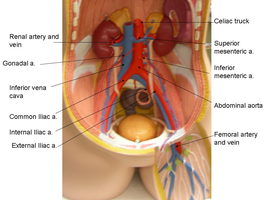
The abdominal cavity contains several major arteries and veins that supply blood to and drain blood from the abdominal organs, pelvic region, and lower limbs. Understanding the anatomy of these vessels is essential for comprehending the circulatory pathways and their clinical significance.
Abdominal aorta: The main arterial trunk supplying oxygenated blood to the abdominal organs and lower body.
Inferior vena cava: The large vein that returns deoxygenated blood from the lower body to the heart.
Renal arteries and veins: Vessels that supply the kidneys and drain filtered blood.
Common iliac arteries and veins: Branches of the aorta and vena cava that supply and drain the pelvis and lower limbs.
Other major branches: Celiac trunk, superior and inferior mesenteric arteries, gonadal arteries, and femoral arteries.
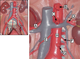
Key Arterial Branches of the Abdominal Aorta
The abdominal aorta gives rise to several important branches that supply the digestive organs, kidneys, and lower limbs.
Celiac trunk: Supplies the stomach, liver, spleen, and upper duodenum.
Superior mesenteric artery: Supplies the small intestine and part of the large intestine.
Inferior mesenteric artery: Supplies the distal large intestine.
Renal arteries: Supply the kidneys.
Gonadal arteries: Supply the testes or ovaries.
Common iliac arteries: Terminal branches of the aorta that supply the pelvis and lower limbs.
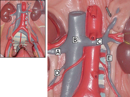
Major Veins of the Abdomen
The inferior vena cava is the principal vein of the abdomen, collecting blood from the lower limbs, pelvis, and abdominal organs.
Inferior vena cava: Formed by the union of the common iliac veins; ascends to the right of the aorta.
Renal veins: Drain the kidneys into the inferior vena cava.
Gonadal veins: Drain the testes or ovaries; right gonadal vein drains directly into the inferior vena cava, left into the left renal vein.
Common iliac veins: Formed by the union of internal and external iliac veins; join to form the inferior vena cava.
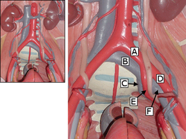
Common Iliac Arteries and Veins
The common iliac arteries and veins are major vessels that bifurcate to supply and drain the pelvis and lower limbs.
Common iliac artery: Divides into internal and external iliac arteries.
Internal iliac artery: Supplies pelvic organs.
External iliac artery: Continues as the femoral artery to supply the lower limb.
Common iliac vein: Formed by the union of internal and external iliac veins; drains into the inferior vena cava.
Summary Table: Major Abdominal Arteries and Veins
The following table summarizes the main arteries and veins of the abdomen, their origins, and the regions they supply or drain.
Vessel | Origin/Termination | Region Supplied/Drained |
|---|---|---|
Abdominal aorta | Continuation of thoracic aorta | Abdominal organs, pelvis, lower limbs |
Celiac trunk | Abdominal aorta | Stomach, liver, spleen, upper duodenum |
Superior mesenteric artery | Abdominal aorta | Small intestine, proximal large intestine |
Inferior mesenteric artery | Abdominal aorta | Distal large intestine |
Renal arteries | Abdominal aorta | Kidneys |
Inferior vena cava | Union of common iliac veins | Lower limbs, pelvis, abdominal organs |
Renal veins | Kidneys | Inferior vena cava |
Common iliac arteries/veins | Terminal branches of aorta/vena cava | Pelvis, lower limbs |
Clinical Relevance
Aneurysms: The abdominal aorta is a common site for aneurysm formation, which can be life-threatening if ruptured.
Renal artery stenosis: Narrowing of the renal arteries can lead to hypertension and kidney damage.
Deep vein thrombosis (DVT): Clots in the iliac or femoral veins can travel to the lungs, causing pulmonary embolism.
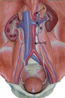
Additional info: The images provided reinforce the anatomical relationships and branching patterns of the major abdominal vessels, which are essential for understanding both normal physiology and clinical pathologies.
