 Back
BackMajor Blood Vessels of the Abdominopelvic Region and Blood Vessel Diseases
Study Guide - Smart Notes
 Tailored notes based on your materials, expanded with key definitions, examples, and context.
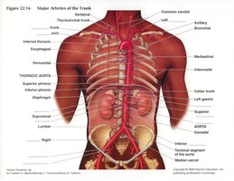
Major Arteries of the Abdominopelvic Region
Overview of the Arterial System
The arterial system of the abdominopelvic region supplies oxygenated blood to the organs of the abdomen and pelvis. Major arteries branch from the aorta and further divide to supply specific organs and tissues.
Aorta: The main artery that descends from the heart and branches into the abdominal aorta, which supplies the lower body.
Brachiocephalic, Subclavian, and Common Iliac Arteries: Major branches that supply the upper limbs, head, and lower limbs, respectively.
Renal Arteries: Supply the kidneys.
Mesenteric Arteries (Superior and Inferior): Supply the intestines.
Splenic and Common Hepatic Arteries: Supply the spleen and liver, respectively.
Arteries Supplying the Abdominopelvic Organs
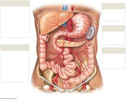
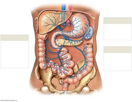
The abdominal aorta gives rise to several branches that supply the digestive organs, spleen, pancreas, and pelvic structures. The celiac trunk, superior mesenteric artery, and inferior mesenteric artery are the primary sources for these regions.
Celiac Trunk: Branches into the left gastric, splenic, and common hepatic arteries, supplying the stomach, spleen, liver, and pancreas.
Superior Mesenteric Artery: Supplies the small intestine and part of the large intestine.
Inferior Mesenteric Artery: Supplies the distal part of the large intestine and rectum.
Other Branches: Include the right and left colic, ileocolic, and sigmoidal arteries, which supply specific segments of the colon.
Major Veins of the Abdominopelvic Region
Venous Drainage of the Abdomen and Chest
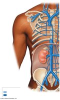
Venous blood from the abdominopelvic organs is collected by a network of veins that ultimately drain into the inferior vena cava. These veins are classified as superficial or deep, depending on their location relative to the body wall.
Inferior Vena Cava: The main vein that returns deoxygenated blood from the lower body to the heart.
Renal, Hepatic, and Gonadal Veins: Drain the kidneys, liver, and gonads, respectively.
Common, Internal, and External Iliac Veins: Drain the pelvis and lower limbs.
Other Veins: Include lumbar, mediastinal, and esophageal veins.
Venous Drainage Pathways
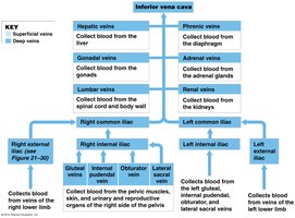
The venous system of the abdomen is organized into a hierarchy, with blood from various organs converging into larger veins before reaching the inferior vena cava.
Vein | Region Drained |
|---|---|
Hepatic veins | Liver |
Renal veins | Kidneys |
Gonadal veins | Gonads |
Lumbar veins | Posterior abdominal wall |
Common iliac veins | Lower limbs and pelvis |
The Hepatic Portal System
Structure and Function
The hepatic portal system is a specialized venous network that directs blood from the gastrointestinal tract and spleen to the liver before it enters the systemic circulation. This allows the liver to process nutrients and detoxify substances absorbed from the digestive tract.
Hepatic Portal Vein: Formed by the union of the superior mesenteric and splenic veins; carries nutrient-rich blood to the liver.
Tributaries: Include the gastric, pancreatic, colic, and rectal veins.
Significance: Ensures that substances absorbed from the intestines are metabolized or detoxified by the liver before reaching the rest of the body.
Blood Vessel Diseases
Common Diseases Affecting Blood Vessels
Blood vessels are susceptible to several diseases that can compromise blood flow and organ function. The most significant conditions include arteriosclerosis, atherosclerosis, and coronary artery disease.
Arteriosclerosis: Hardening and loss of elasticity of arterial walls, often due to aging.
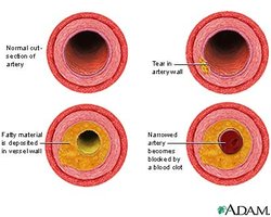
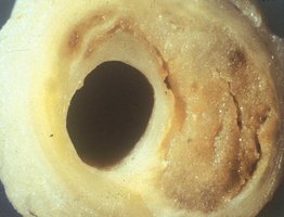
Atherosclerosis: Accumulation of lipoprotein-rich plaques within arterial walls, leading to narrowing and reduced blood flow.
Coronary Artery Disease: Atherosclerosis specifically affecting the coronary arteries, which supply the heart muscle.
Pathophysiology: Plaque buildup can lead to vessel narrowing, reduced tissue perfusion, and increased risk of clot formation, which may result in heart attack or stroke.
Summary Table: Blood Vessel Diseases
Disease | Description | Main Consequence |
|---|---|---|
Arteriosclerosis | Hardening of arterial walls | Reduced elasticity, increased blood pressure |
Atherosclerosis | Plaque buildup inside arteries | Narrowed vessels, risk of clot |
Coronary Artery Disease | Atherosclerosis of coronary arteries | Reduced blood flow to heart, risk of heart attack |
Key Terms and Concepts
Artery: Blood vessel that carries blood away from the heart.
Vein: Blood vessel that returns blood to the heart.
Hepatic Portal System: Venous system directing blood from the digestive tract to the liver.
Plaque: Fatty deposit within the wall of a blood vessel.
Additional info:
Understanding the anatomy of the major blood vessels and their diseases is essential for comprehending how blood is distributed and regulated throughout the body, and how vascular diseases can impact systemic health.
