 Back
BackMajor Muscles of the Human Body: Structure and Function
Study Guide - Smart Notes
 Tailored notes based on your materials, expanded with key definitions, examples, and context.
Muscles of the Head
Facial Muscles and Their Functions
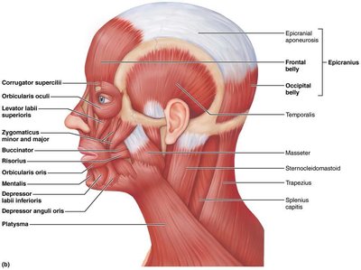
The muscles of the human head are primarily responsible for facial expression, mastication (chewing), and movements of the scalp. These muscles are unique because many insert into the skin rather than bone, allowing for a wide range of expressive movements.
Epicranius (Frontal and Occipital Belly): The frontal belly raises the eyebrows, while the occipital belly fixes the aponeurosis and pulls the scalp posteriorly.
Orbicularis oculi: Closes the eyelids, essential for blinking and protecting the eye.
Zygomaticus (Major and Minor): Raises the lateral corners of the mouth, as in smiling.
Risorius: Pulls the corner of the lip laterally, contributing to expressions such as grinning.
Orbicularis oris: Closes and protrudes the lips, as in kissing or whistling.
Buccinator: Compresses the cheek, important in sucking and blowing.
Muscles of Mastication
These muscles are responsible for movements of the jaw during chewing and speaking.
Masseter: Prime mover of jaw closure, elevates the mandible.
Temporalis: Elevates and retracts the mandible, assisting in jaw closure.
Buccinator: Assists in mastication by keeping food between the teeth.
Muscles of the Neck
Anterolateral Muscles of the Neck
The neck muscles facilitate head movement, swallowing, and breathing. They also provide structural support for the head.
Platysma: Tenses the skin of the neck and helps depress the mandible.
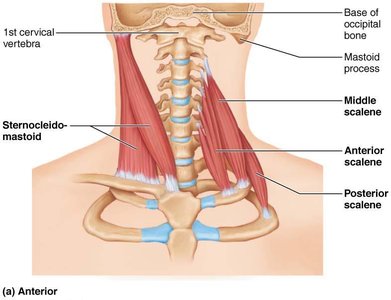
Sternocleidomastoid: Flexes and laterally rotates the head; important for head rotation and flexion.
Scalenes (anterior, middle, posterior): Elevate the first two ribs during inspiration and assist in neck flexion.
Muscles of the Thorax and Shoulder
Superficial Muscles
These muscles are involved in movements of the shoulder and upper arm, as well as respiration.
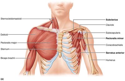
Pectoralis major: Adducts and medially rotates the arm.
Pectoralis minor: With ribs fixed, pulls the scapula forward and downward.
Deltoid: Prime mover of arm abduction when all fibers contract simultaneously.
Serratus anterior: Rotates the scapula so its inferior angle moves laterally and upward.
Deep Muscles of the Thorax
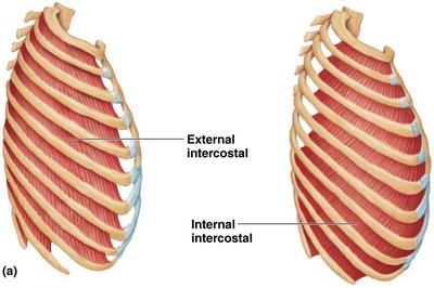
These muscles are essential for the mechanics of breathing, controlling the movement of the rib cage and diaphragm.
External intercostals: Pull ribs toward one another to elevate the rib cage during inspiration.
Internal intercostals: Pull ribs together and depress the rib cage during forced expiration.
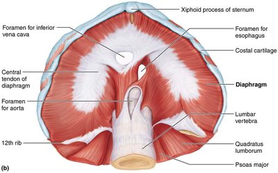
Diaphragm: Prime mover of inspiration; flattens on contraction to increase thoracic cavity volume.
Muscles of the Abdominal Wall
Structure and Function
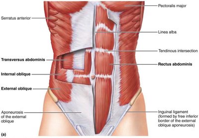
The abdominal wall muscles support the trunk, allow movement, and help maintain posture. They also protect internal organs and assist in processes such as forced expiration, urination, and childbirth.
Rectus abdominis: Flexes and rotates the lumbar region of the vertebral column.
External oblique: Flexes the vertebral column and compresses the abdominal wall.
Internal oblique: Same function as the external oblique; acts synergistically.
Transversus abdominis: Compresses abdominal contents.
Posterior Muscles of the Trunk
Muscles of the Neck, Shoulder, and Thorax
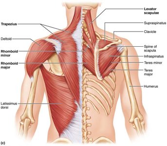
These muscles stabilize and move the scapula and upper limb, and are important for posture and upper body movement.
Trapezius: Stabilizes, elevates, retracts, and rotates the scapula.
Supraspinatus: Initiates abduction of the arm.
Infraspinatus: Rotates the arm laterally.
Teres major: Extends, medially rotates, and adducts the arm.
Teres minor: Same as infraspinatus.
Rhomboideus major and minor: Stabilize the scapula.
Levator scapulae: Elevates and adducts the scapula.
Latissimus dorsi: Prime mover of arm extension, powerful arm adductor, medially rotates the arm.
Muscles That Act on the Forearm
Anterior and Posterior Compartments
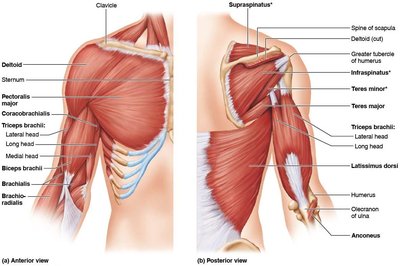
These muscles are responsible for flexion, extension, and rotation of the forearm at the elbow joint.
Brachioradialis: Synergist in flexing the forearm.
Triceps brachii: Powerful forearm extensor.
Anconeus: Synergist of triceps brachii in elbow extension.
Biceps brachii: Flexes and supinates the forearm.
Brachialis: Major forearm flexor.
Muscles That Act on the Hand and Fingers
Anterior Compartment (Flexors)
These muscles primarily flex the wrist and fingers, allowing for gripping and fine motor movements.
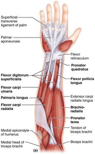
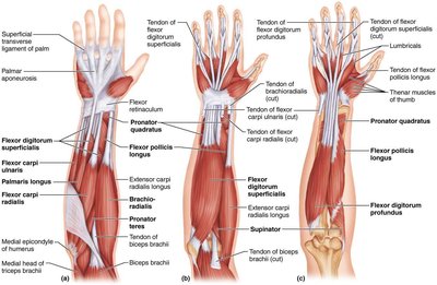
Pronator teres: Pronates the forearm.
Flexor carpi radialis: Powerful flexor of the hand.
Palmaris longus: Tenses the fascia of the palm during hand movements.
Flexor carpi ulnaris: Powerful flexor and adductor of the hand.
Flexor digitorum superficialis: Flexes the hand and middle phalanges of the second to fifth fingers.
Posterior Compartment (Extensors)
These muscles extend the wrist and fingers, essential for releasing objects and hand positioning.
Extensor carpi radialis (longus and brevis): Extends and abducts the hand.
Extensor digitorum: Prime mover of finger extension.
Extensor carpi ulnaris: Extends and adducts the hand.
Muscles of the Pelvis and Thigh
Origin of the Pelvis
These muscles are responsible for movements of the thigh at the hip joint, including flexion, extension, abduction, and rotation.
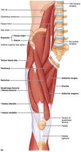
Iliopsoas (Iliacus and Psoas major): Prime mover for flexing the thigh or trunk on the thigh; psoas major also flexes the vertebral column laterally.
Sartorius: Flexes, abducts, and laterally rotates the thigh.
Medial Compartment of the Thigh
These muscles primarily adduct the thigh and assist in flexion and medial rotation.
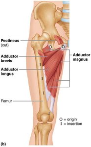
Adductor magnus, longus, brevis: Adduct, flex, and medially rotate the thigh.
Pectineus: Adducts, flexes, and medially rotates the thigh.
Gracilis: Adducts the thigh, flexes, and medially rotates the leg.
Anterior Compartment of the Thigh
These muscles extend the leg at the knee and flex the thigh at the hip.
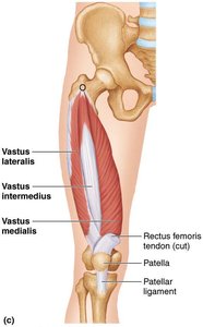
Quadriceps femoris (Rectus femoris, Vastus lateralis, Vastus medialis, Vastus intermedius): Extend the leg; rectus femoris also flexes the thigh at the hip.
Tensor fasciae latae: Strengthens the leg and trunk on the thigh.
Gluteal Region
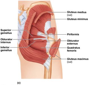
The gluteal muscles are responsible for extension, abduction, and rotation of the thigh.
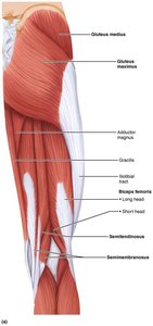
Gluteus maximus: Major extensor of the thigh.
Gluteus medius: Abducts and medially rotates the thigh.
Posterior Compartment (Hamstrings)
The hamstring muscles extend the thigh and flex the leg at the knee joint.
Biceps femoris: Extends the thigh and flexes the leg.
Semitendinosus: Extends the thigh and flexes the leg.
Semimembranosus: Extends the thigh and flexes the leg.
Muscles of the Leg
Compartments and Main Actions
The muscles of the leg are divided into anterior, lateral, and posterior compartments, each responsible for specific movements of the foot and toes.
Lateral compartment (Fibularis longus and brevis): Plantar flexes and everts the foot.
Anterior compartment (Tibialis anterior, Extensor digitorum longus): Tibialis anterior is the prime mover of dorsiflexion; extensor digitorum longus is the prime mover of toe extension.
Posterior compartment (Triceps surae: Gastrocnemius and Soleus): Plantar flexes the foot.
Example: The quadriceps femoris group is essential for standing up from a seated position, while the hamstrings are crucial for walking and running.
Additional info: The arrangement of muscles into compartments allows for efficient movement and coordination, with each compartment typically innervated by specific nerves and supplied by distinct blood vessels.
