 Back
Male and Female Reproductive Anatomy & Histology Study Guide
Study Guide - Smart Notes
 Tailored notes based on your materials, expanded with key definitions, examples, and context.
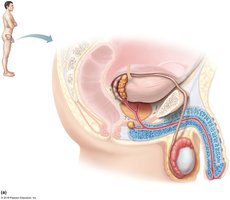
Q1. Label the male reproductive structures on the image below.
Background
Topic: Male Reproductive Anatomy
This question tests your knowledge of the anatomical structures of the male reproductive system, including organs involved in sperm production, maturation, and delivery.
Key Terms:
Testis: Site of sperm production.
Epididymis: Sperm maturation and storage.
Vas deferens: Transports sperm from epididymis to urethra.
Seminal vesicle, prostate gland: Contribute fluids to semen.
Urethra: Passageway for urine and semen.
Penis: External organ for delivery of semen.
Step-by-Step Guidance
Identify the testis in the scrotum, which is the site of sperm production.
Locate the epididymis, which sits atop the testis and is where sperm mature.
Trace the vas deferens as it leaves the epididymis and travels upward toward the pelvic cavity.
Find the seminal vesicle and prostate gland, which add fluids to the sperm to form semen.
Follow the urethra as it passes through the penis, serving as the final pathway for semen delivery.
Try solving on your own before revealing the answer!
Final Answer:
The labeled structures are: testis, epididymis, vas deferens, seminal vesicle, prostate gland, urethra, and penis.
Each structure plays a specific role in sperm production, maturation, and delivery.
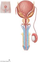
Q2. Label the male reproductive structures on the image below.
Background
Topic: Male Reproductive Anatomy (Frontal View)
This question focuses on identifying the male reproductive organs from a frontal perspective, emphasizing the spatial relationships between the bladder, prostate, and penis.
Key Terms:
Bladder: Stores urine.
Prostate gland: Adds fluid to semen.
Urethra: Runs through the penis, carrying urine and semen.
Penis: External organ for semen delivery.
Testis and epididymis: Sperm production and maturation.
Step-by-Step Guidance
Identify the bladder at the top of the image, which stores urine.
Locate the prostate gland beneath the bladder.
Trace the urethra as it passes through the prostate and penis.
Find the testis and epididymis on the side, connected to the vas deferens.
Try solving on your own before revealing the answer!
Final Answer:
The labeled structures are: bladder, prostate gland, urethra, penis, testis, and epididymis.
Understanding their spatial arrangement helps clarify their functions in reproduction and urination.
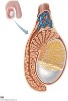
Q3. Label the male reproductive structures on the image below.
Background
Topic: Testicular Anatomy
This question tests your ability to identify internal structures of the testis, including those involved in sperm production and transport.
Key Terms:
Seminiferous tubules: Site of sperm production.
Rete testis: Network connecting seminiferous tubules to epididymis.
Epididymis: Sperm maturation and storage.
Blood vessels: Supply nutrients and remove waste.
Step-by-Step Guidance
Identify the seminiferous tubules, which are coiled structures inside the testis.
Locate the rete testis, which connects the seminiferous tubules to the epididymis.
Find the epididymis, which wraps around the testis and stores sperm.
Observe the blood vessels at the top, which support testicular function.
Try solving on your own before revealing the answer!
Final Answer:
The labeled structures are: seminiferous tubules, rete testis, epididymis, and blood vessels.
These structures are essential for sperm production, maturation, and transport.
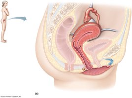
Q4. Label the female reproductive structures on the image below.
Background
Topic: Female Reproductive Anatomy (Sagittal View)
This question tests your knowledge of the internal female reproductive organs, including the uterus, vagina, and associated structures.
Key Terms:
Uterus: Site of implantation and fetal development.
Vagina: Birth canal and site of sperm entry.
Bladder: Stores urine.
Rectum: Part of digestive system, located posteriorly.
Step-by-Step Guidance
Identify the uterus in the center, which is pear-shaped.
Locate the vagina below the uterus, leading to the external genitalia.
Find the bladder anterior to the uterus.
Observe the rectum posterior to the uterus.
Try solving on your own before revealing the answer!
Final Answer:
The labeled structures are: uterus, vagina, bladder, and rectum.
These organs are key to reproductive and urinary functions.
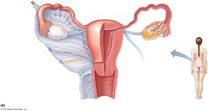
Q5. Label the female reproductive structures on the image below.
Background
Topic: Female Reproductive Anatomy (Frontal View)
This question focuses on the spatial arrangement of the ovaries, fallopian tubes, uterus, and associated structures.
Key Terms:
Ovary: Produces eggs and hormones.
Fallopian tube: Transports eggs to uterus.
Uterus: Site of implantation and fetal development.
Cervix: Lower part of uterus, opening to vagina.
Step-by-Step Guidance
Identify the ovaries on each side, which produce eggs.
Locate the fallopian tubes connecting the ovaries to the uterus.
Find the uterus in the center, with the cervix at its lower end.
Try solving on your own before revealing the answer!
Final Answer:
The labeled structures are: ovary, fallopian tube, uterus, and cervix.
These structures are essential for egg transport, fertilization, and fetal development.
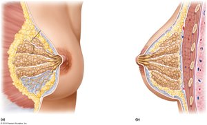
Q6. Label the female reproductive structures on the image below.
Background
Topic: Mammary Gland Anatomy
This question tests your understanding of the internal structure of the mammary gland, including lobes, ducts, and the lactiferous sinus.
Key Terms:
Lobes: Glandular tissue producing milk.
Lactiferous ducts: Transport milk from lobes to nipple.
Lactiferous sinus: Storage area for milk before release.
Areola: Pigmented area around nipple.
Step-by-Step Guidance
Identify the lobes, which are clusters of glandular tissue.
Locate the lactiferous ducts, which carry milk toward the nipple.
Find the lactiferous sinus, which stores milk before it is released.
Observe the areola and nipple, which are external landmarks.
Try solving on your own before revealing the answer!
Final Answer:
The labeled structures are: lobes, lactiferous ducts, lactiferous sinus, areola, and nipple.
These components are crucial for milk production and delivery.
