 Back
BackMechanisms of Digestion and Absorption: A Mini-Textbook Study Guide
Study Guide - Smart Notes
 Tailored notes based on your materials, expanded with key definitions, examples, and context.
Mechanisms of Digestion and Absorption
Overview of Digestion and Absorption
Digestion is the catabolic process by which ingested food is broken down into its chemical building blocks, allowing for absorption into the body. Only molecules small enough to cross the wall of the small intestine can be absorbed. Enzymatic hydrolysis, primarily by intrinsic and accessory gland enzymes, is the main mechanism of digestion, where water is added to break chemical bonds in macromolecules.
Digestion: Breaks down macromolecules (carbohydrates, proteins, fats) into monomers (monosaccharides, amino acids, fatty acids, and glycerol).
Absorption: The process of moving digested end products from the lumen of the gut into the body, primarily through the epithelial cells of the small intestine.
Tight junctions: Ensure that substances must pass through, not between, epithelial cells.
Lipid absorption: Lipids can diffuse passively, but most other nutrients require active transport mechanisms.
Site of absorption: Most nutrients are absorbed before chyme reaches the ileum.
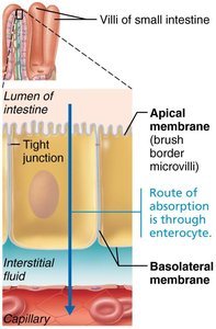
Digestion and Absorption of Carbohydrates
Enzymatic Breakdown and Absorption Pathways
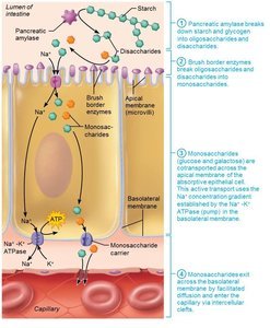
Carbohydrate digestion begins in the mouth and continues in the small intestine. Only monosaccharides (glucose, fructose, galactose) can be absorbed. Starch and disaccharides are broken down into oligosaccharides and disaccharides, then further into monosaccharides by brush border enzymes.
Salivary amylase: Begins starch digestion in the mouth.
Pancreatic amylase: Continues digestion in the small intestine, breaking down starch and glycogen into oligosaccharides and disaccharides.
Brush border enzymes: Dextrinase, lactase, glucoamylase, maltase, and sucrase complete the breakdown into monosaccharides.
Absorption: Monosaccharides are absorbed via secondary active transport (with Na+) or facilitated diffusion across the apical membrane, then exit via facilitated diffusion across the basolateral membrane into the blood.
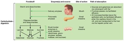
Mechanism of Carbohydrate Absorption
Monosaccharides are transported across the intestinal epithelium by specific mechanisms:
Glucose and galactose: Cotransported with Na+ via secondary active transport (SGLT1 transporter).
Fructose: Absorbed by facilitated diffusion (GLUT5 transporter).
All monosaccharides: Exit the basolateral membrane by facilitated diffusion (GLUT2 transporter) and enter the capillary blood in the villi.
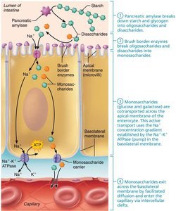
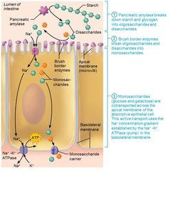
Clinical Correlation: Lactose Intolerance
Lactose intolerance results from a deficiency of the enzyme lactase, leading to the inability to digest lactose. Undigested lactose creates an osmotic gradient, preventing water absorption and causing diarrhea. Bacterial fermentation of lactose produces gas, leading to bloating, flatulence, and cramping. Treatment involves lactase supplements or avoiding lactose-containing foods.
Digestion and Absorption of Proteins
Enzymatic Breakdown and Absorption Pathways
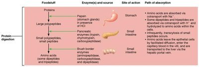
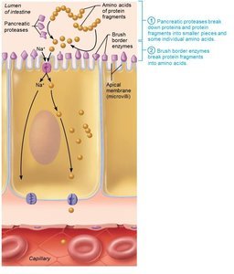
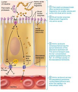
Protein digestion begins in the stomach and continues in the small intestine. Proteins are broken down into large polypeptides, then into smaller peptides and amino acids by various enzymes.
Stomach: Pepsin (active at pH 1.5–2.5) initiates protein digestion, producing large polypeptides.
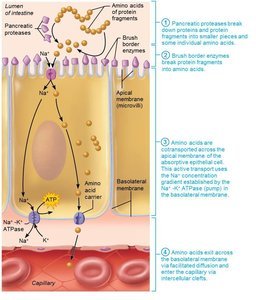
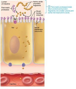
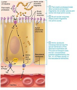
Small intestine: Pancreatic proteases (trypsin, chymotrypsin, carboxypeptidase) and brush border enzymes (aminopeptidases, carboxypeptidases, dipeptidases) further break down peptides into amino acids.
Absorption: Amino acids are absorbed via secondary active transport (with Na+ or H+) across the apical membrane and exit via facilitated diffusion across the basolateral membrane.
Mechanism of Protein Absorption
Amino acids and small peptides are transported across the intestinal epithelium by specific mechanisms:
Amino acids: Cotransported with Na+ or H+ via secondary active transporters.
Dipeptides and tripeptides: Absorbed by H+-dependent cotransporters and hydrolyzed to amino acids within epithelial cells.
All amino acids: Exit the basolateral membrane by facilitated diffusion and enter the capillary blood in the villi.
Summary Table: Digestion and Absorption of Carbohydrates and Proteins
Foodstuff | Enzyme(s) and Source | Site of Action | Path of Absorption |
|---|---|---|---|
Starch and Disaccharides | Salivary amylase (mouth), Pancreatic amylase (small intestine), Brush border enzymes (small intestine) | Mouth, Small intestine | Glucose and galactose: cotransport with Na+; Fructose: facilitated diffusion; All exit via facilitated diffusion to blood |
Proteins | Pepsin (stomach), Pancreatic proteases (small intestine), Brush border enzymes (small intestine) | Stomach, Small intestine | Amino acids: cotransport with Na+ or H+; Dipeptides/tripeptides: cotransport with H+; All exit via facilitated diffusion to blood |
Additional info: The mechanisms described here are essential for understanding nutrient assimilation and the consequences of malabsorption syndromes, such as lactose intolerance or protein-energy malnutrition.
Digestion and Absorption of Lipids
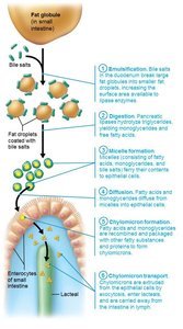
Overview of Lipid Digestion
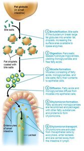
Lipid digestion is a complex process that occurs primarily in the small intestine, involving several steps to break down insoluble triglycerides into absorbable molecules.
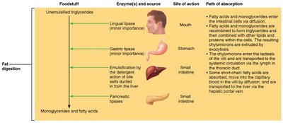
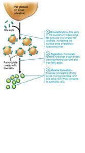
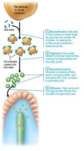
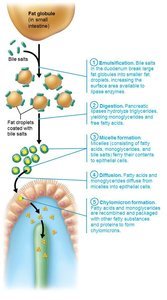
Emulsification: Bile salts break large fat globules into smaller droplets, increasing surface area for enzyme action.
Digestion: Pancreatic lipases hydrolyze triglycerides into monoglycerides and free fatty acids.
Micelle Formation: Digestion products are coated with bile salts and lecithin, forming micelles that facilitate transport across the intestinal epithelium.
Diffusion: Lipid products leave micelles and diffuse across the epithelial membrane.
Chylomicron Formation: Lipid products are reassembled into triglycerides and packaged with proteins to form chylomicrons.
Chylomicron Transport: Chylomicrons are exocytosed into lymphatic lacteals and eventually enter venous blood.
Stepwise Process of Fat Digestion and Absorption
Emulsification: Bile salts break up fat globules.
Digestion: Pancreatic lipases hydrolyze triglycerides.
Micelle Formation: Bile salts and lecithin form micelles with digestion products.
Diffusion: Fatty acids and monoglycerides diffuse into epithelial cells.
Chylomicron Formation: Lipid products are reassembled and packaged.
Chylomicron Transport: Chylomicrons enter lymphatic lacteals.
Digestion and Absorption of Nucleic Acids
Overview of Nucleic Acid Digestion
Nucleic acids from ingested cells are broken down in the small intestine by specific enzymes.
Pancreatic Nucleases: Hydrolyze DNA and RNA into nucleotide monomers.
Brush Border Enzymes: Nucleosidases and phosphatases further break down nucleotides into nitrogenous bases, pentose sugars, and phosphate ions.
Absorption: Breakdown products are actively transported by carriers in the epithelium of villi.
Absorption of Vitamins, Electrolytes, and Water
Vitamin Absorption
Fat-soluble vitamins (A, D, E, K): Carried by micelles and diffuse into absorptive cells.
Water-soluble vitamins (C, B): Absorbed by diffusion or via passive/active transporters.
Vitamin B12: Binds with intrinsic factor and is absorbed by endocytosis.
Large intestine: Absorbs vitamin K and B vitamins produced by bacterial metabolism.
Electrolyte Absorption
Most ions are actively transported along the small intestine.
Iron and calcium are absorbed in the duodenum; iron is stored with ferritin and transported by transferrin when needed.
Calcium absorption is regulated by vitamin D and parathyroid hormone (PTH).
Sodium absorption is coupled with glucose and amino acid absorption.
Potassium diffuses in response to osmotic gradients.
Water Absorption
About 9 liters of water enter the small intestine daily, mostly from GI tract secretions.
95% is absorbed in the small intestine by osmosis; most of the rest is absorbed in the large intestine.
Water uptake is coupled with solute uptake.
Clinical Aspects and Homeostatic Imbalances
Malabsorption Syndromes
Caused by anything interfering with delivery of bile or pancreatic juice.
Damaged intestinal mucosa (e.g., bacterial infection, antibiotics) can impair absorption.
Celiac Disease: Immune reaction to gluten damages intestinal villi and brush border; treatment is gluten-free diet.
Congenital and Genetic Disorders
Cleft Palate and Cleft Lip: Failure of palatine bones/processes to fuse; affects feeding in infants.
Tracheoesophageal Fistula: Abnormal connection between esophagus and trachea.
Cystic Fibrosis: Thick mucus blocks pancreatic duct, affecting digestion.
Developmental Aspects of the Digestive System
Embryonic Development
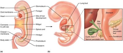
The digestive system develops from the primitive gut, with the epithelial lining formed from endoderm and other structures from mesoderm.
Oral membrane: Becomes the mouth.
Cloacal membrane: Becomes the anus.
By week 5, the alimentary canal is a continuous tube from mouth to anus.
Accessory organs bud from the mucosa shortly after.
Digestive System After Birth
Fetal nutrition occurs via placenta; GI tract matures by swallowing amniotic fluid.
Newborns have rooting and sucking reflexes for feeding.
Newborns double birth weight in 6 months; adult diet by 2 years.
Gastroenteritis, cholecystitis, and ulcers are common in middle age.
Aging and the Digestive System
GI tract activity declines with age; less digestive juice, less efficient absorption, slower peristalsis.
Taste and smell decline; periodontal disease is common.
Diverticulosis, fecal incontinence, and GI cancers are frequent in elderly.
Stomach and colon cancers rarely have early symptoms; metastasized colon cancer often spreads to liver.
Oral cancers can be detected during dental exams; most GI cancers are treatable if caught early.
Prevention: regular dental and medical examinations.
