Back
BackMembrane Potentials, Action Potentials, and Synaptic Transmission in Neurons
Study Guide - Smart Notes
 Tailored notes based on your materials, expanded with key definitions, examples, and context.
Resting Membrane Potential
Definition and Ionic Basis
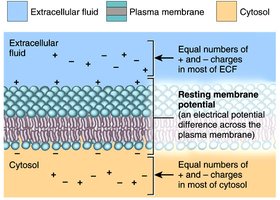
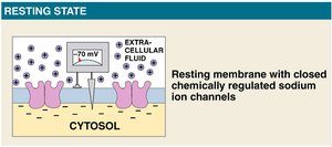
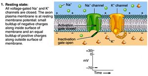
The resting membrane potential (RMP) is the electrical potential difference across the plasma membrane of a resting cell, typically measured at -70 mV in neurons. This potential is established by differences in ion concentrations and the selective permeability of the membrane to these ions.
Transmembrane potential: The voltage difference between the inside and outside of the cell.
Na+/K+ pump: Actively transports 3 Na+ ions out and 2 K+ ions into the cell, contributing to the negative charge inside.
Charge separation: The cytosol and extracellular fluid each have equal numbers of positive and negative charges, but the membrane itself maintains a small charge separation.
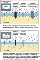
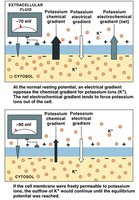
Electrochemical Gradients
The movement of ions across the membrane is driven by both chemical (concentration) and electrical (voltage) gradients, collectively called the electrochemical gradient.
Sodium (Na+): The equilibrium potential for Na+ is +66 mV. At rest, both chemical and electrical gradients drive Na+ into the cell.
Potassium (K+): The equilibrium potential for K+ is -90 mV. The chemical gradient drives K+ out, but the electrical gradient opposes this movement.
Gated Channels and Changes in Transmembrane Potential
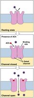
Chemically-Regulated Channels
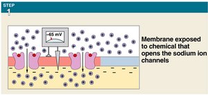
Chemically-regulated (ligand-gated) channels open or close in response to the binding of specific chemicals, such as neurotransmitters (e.g., acetylcholine, ACh). These channels are primarily found on postsynaptic membranes, including dendrites and cell bodies of neurons.
Opening these channels can lead to local changes in membrane potential.
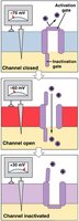
Voltage-Regulated Channels
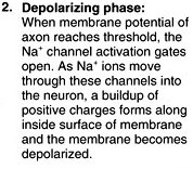
Voltage-regulated (voltage-gated) channels open or close in response to changes in the transmembrane potential. They are abundant along excitable membranes, such as axons and muscle fiber sarcolemma.
Types include voltage-gated Na+, K+, and Ca2+ channels.
Graded Potentials
Characteristics and Types
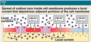
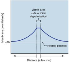
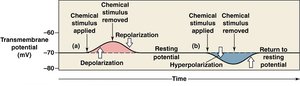
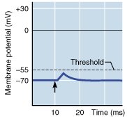
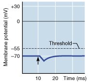
Graded potentials are local, short-lived changes in the membrane potential that can be depolarizing or hyperpolarizing. They result from the opening of gated ion channels and are not propagated over long distances.
Depolarization: Membrane potential becomes less negative (more positive), usually due to Na+ influx.
Hyperpolarization: Membrane potential becomes more negative than resting, often due to K+ efflux.
Postsynaptic Potentials and Summation
Excitatory and Inhibitory Postsynaptic Potentials
Postsynaptic potentials (PSPs) are graded potentials that occur on the postsynaptic membrane in response to neurotransmitters.
Excitatory postsynaptic potential (EPSP): Graded depolarization, often due to Na+ influx.
Inhibitory postsynaptic potential (IPSP): Graded hyperpolarization, often due to K+ efflux.
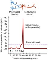
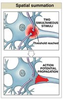
Summation: Temporal and Spatial
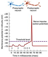
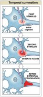
Summation is the process by which multiple graded potentials combine to influence the likelihood of an action potential.
Temporal summation: Rapid succession of stimuli at a single synapse.
Spatial summation: Simultaneous stimuli at multiple synapses.
Action Potentials
Generation and Propagation
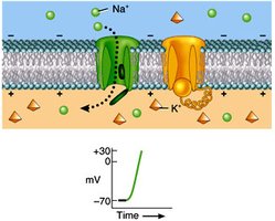
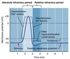
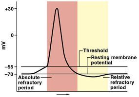
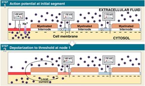
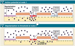
Action potentials are rapid, large changes in membrane potential that propagate along excitable membranes. They are all-or-none events triggered when the membrane reaches threshold (typically -60 to -55 mV).
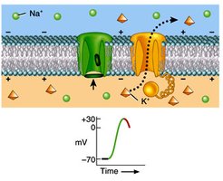
Depolarization: Voltage-gated Na+ channels open, Na+ enters, membrane potential rises to +30 mV.
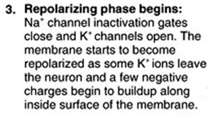
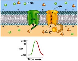
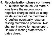
Repolarization: Na+ channels inactivate, K+ channels open, K+ exits, membrane potential returns toward -70 mV.
Hyperpolarization: K+ outflow may briefly make the membrane more negative than resting potential.
Na+/K+ pump: Restores original ion concentrations.
Refractory Periods
The refractory period is the time during which a neuron is less sensitive or completely insensitive to further stimulation.
Absolute refractory period: No action potential can be generated (Na+ channels open or inactivated).
Relative refractory period: A stronger-than-normal stimulus can trigger another action potential (K+ channels open, membrane hyperpolarized).
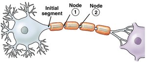
Conduction of Action Potentials
Continuous vs. Saltatory Conduction
Action potentials propagate differently in myelinated and unmyelinated axons.
Continuous conduction: Occurs in unmyelinated axons; action potential moves smoothly along the membrane.
Saltatory conduction: Occurs in myelinated axons; action potential "jumps" from node to node (Nodes of Ranvier), increasing speed.
Synaptic Transmission
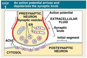
Mechanism at a Cholinergic Synapse
Synaptic transmission is the process by which a neuron communicates with another cell at a synapse, often using neurotransmitters such as acetylcholine (ACh).
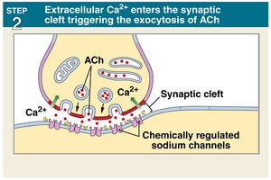
Action potential arrives at the synaptic terminal, opening voltage-gated Ca2+ channels.
Ca2+ influx triggers exocytosis of ACh into the synaptic cleft.
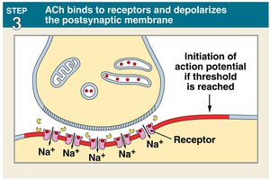
ACh binds to receptors on the postsynaptic membrane, opening chemically-regulated Na+ channels and causing depolarization.
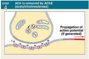
ACh is removed by acetylcholinesterase (AChE), terminating the signal.
Neural Circuits
Types of Neural Circuits
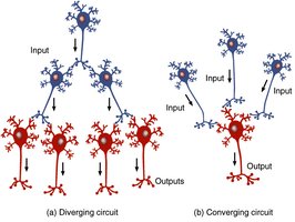
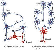
Neuronal pools in the CNS are organized into circuits that determine the flow of information and functional output.
Serial processing: Neurons are arranged in a line, each relaying information to the next.
Divergence: One neuron synapses with several others, spreading the signal.
Convergence: Several neurons synapse on a single neuron, integrating input.
Reverberation: Neurons provide feedback to earlier neurons in the circuit, sustaining activity.
Parallel after-discharge: A single input is relayed along several pathways to a common output.