 Back
BackMembrane Potentials and Action Potentials in Neurons
Study Guide - Smart Notes
 Tailored notes based on your materials, expanded with key definitions, examples, and context.
Membrane Potentials and Neuronal Excitability
Basic Principles of Electricity in Neurons
Neurons, like all cells, possess a resting membrane potential, but they are unique in their ability to rapidly change this potential, making them highly excitable. The movement of ions across the neuronal membrane underlies all electrical signaling in the nervous system.
Opposite charges attract, and energy is required to keep them separated across a membrane.
When separated, the system has potential energy that can be used to do work when charges move toward each other.
Types of Gated Ion Channels
Ion channels are proteins that allow specific ions to cross the membrane. There are three main types of gated channels in neurons:
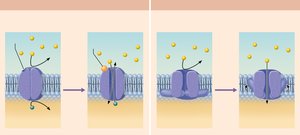
Chemically gated (ligand-gated) channels: Open in response to binding of a specific chemical, such as a neurotransmitter.
Voltage-gated channels: Open or close in response to changes in membrane potential.
Mechanically gated channels: Open or close in response to physical deformation of the receptor (e.g., touch receptors).
Ion Movement and Ohm's Law
When gated channels open, ions diffuse rapidly:
Along chemical gradients (from high to low concentration).
Along electrical gradients (toward opposite charge).
The combined effect is called the electrochemical gradient.
Ion flow creates an electrical current, and voltage changes across the membrane. This relationship is described by Ohm’s law:
Where V is voltage, I is current, and R is resistance.
Resting Membrane Potential
Establishment and Maintenance
The resting membrane potential is typically around -70 mV in neurons. It is established by differences in ion concentrations and membrane permeability:
Na+ concentration is higher outside the cell; K+ concentration is higher inside.
Na+-K+ pumps maintain these gradients by pumping Na+ out and K+ in.
The membrane is more permeable to K+ (due to leakage channels), so K+ leaves the cell, making the inside more negative.
Small Na+ influx slightly offsets the negativity.

Summary Table: Ion Concentrations and Membrane Permeability
Ion | Concentration Outside Cell | Concentration Inside Cell | Relative Permeability |
|---|---|---|---|
Na+ | High (140 mM) | Low (15 mM) | Low |
K+ | Low (5 mM) | High (140 mM) | High |
Changes in Membrane Potential
Types of Changes
Membrane potential changes when ion concentrations or membrane permeability change. These changes produce two types of signals:
Graded potentials: Short-distance, localized changes in membrane potential.
Action potentials: Long-distance signals, especially in axons.
Key terms:
Depolarization: Membrane potential becomes less negative (moves toward zero or above); increases probability of impulse generation.
Hyperpolarization: Membrane potential becomes more negative; decreases probability of impulse generation.
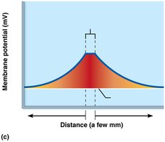
Graded Potentials
Characteristics and Spread
Graded potentials are short-lived, localized changes in membrane potential, triggered by a stimulus that opens gated ion channels. The magnitude depends on stimulus strength and decays with distance.
Receptor potential: Occurs in sensory receptors.
Postsynaptic potential: Occurs in neurons after synaptic transmission.
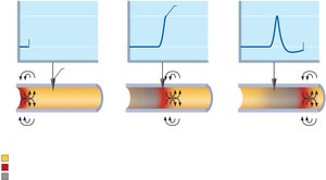
Action Potentials
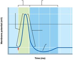
Generation and Phases
An action potential (AP) is the principal way neurons send long-distance signals. It is a brief reversal of membrane potential (~100 mV change) and does not decay with distance. APs occur only in muscle cells and axons of neurons.
Resting state: All gated Na+ and K+ channels are closed.
Depolarization: Na+ channels open, Na+ enters the cell.
Repolarization: Na+ channels inactivate, K+ channels open, K+ exits the cell.
Hyperpolarization: Some K+ channels remain open, Na+ channels reset.
Threshold and All-or-None Principle
Not all depolarizations trigger an AP. The membrane must reach a threshold voltage (typically 15–20 mV above resting) for an AP to occur. If threshold is reached, the AP is generated fully; if not, no AP occurs. This is the all-or-none phenomenon.
Propagation of Action Potentials
APs are propagated along the axon by local currents that depolarize adjacent membrane areas, opening voltage-gated Na+ channels. This process is self-propagating and unidirectional due to channel inactivation behind the AP.
Coding for Stimulus Intensity
All APs are identical in amplitude. The frequency of APs encodes stimulus intensity: higher frequency means a stronger stimulus.
Refractory Periods
After an AP, the neuron cannot immediately fire another AP due to:
Absolute refractory period: No AP possible, regardless of stimulus strength.
Relative refractory period: AP possible only with a stronger-than-usual stimulus.
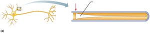
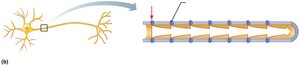
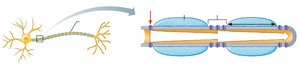
Conduction Velocity
Factors Affecting Speed
AP conduction velocity depends on:
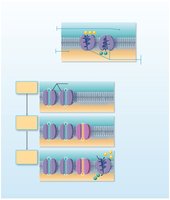
Axon diameter: Larger diameter = faster conduction (less resistance).
Degree of myelination: Myelinated axons conduct faster due to saltatory conduction.
Types of conduction:
Continuous conduction: Slow, occurs in nonmyelinated axons.
Saltatory conduction: Fast, occurs in myelinated axons; APs jump from gap to gap (nodes of Ranvier).
Nerve Fiber Classification
Group | Diameter | Myelination | Conduction Speed | Function |
|---|---|---|---|---|
A | Largest | Myelinated | ~150 m/s | Somatic sensory & motor (skin, muscles, joints) |
B | Intermediate | Lightly myelinated | ~15 m/s | ANS visceral motor & sensory |
C | Smallest | Unmyelinated | ~1 m/s | ANS visceral motor & sensory |
