 Back
BackMembranes: Structure, Types, and Clinical Relevance
Study Guide - Smart Notes
 Tailored notes based on your materials, expanded with key definitions, examples, and context.
Membranes in the Human Body
Overview of Body Membranes
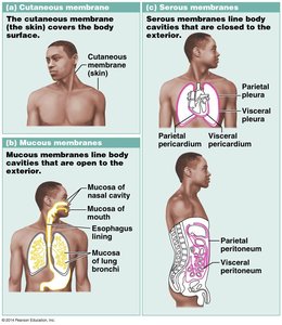
Body membranes are essential anatomical structures composed of epithelial tissue supported by underlying connective tissue. They serve as protective barriers, line body cavities, and facilitate physiological processes such as secretion and absorption. There are three primary types of membranes: cutaneous, mucous, and serous membranes.
Cutaneous membrane: The skin, covering the external surface of the body.
Mucous membranes: Line body cavities that open to the exterior.
Serous membranes: Line closed body cavities and cover organs within these cavities.
Cutaneous Membrane
Structure and Function
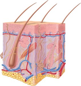
The cutaneous membrane, commonly known as the skin, is a dry membrane that forms the body's outer protective covering. It consists of two main layers:
Epidermis: Composed of keratinized stratified squamous epithelium, providing a tough, water-resistant barrier.
Dermis: Made of dense irregular connective tissue, containing blood vessels, nerves, and accessory structures.
The skin protects against mechanical injury, pathogens, and dehydration, and plays a role in thermoregulation and sensation.
Mucous Membranes
Structure, Locations, and Functions
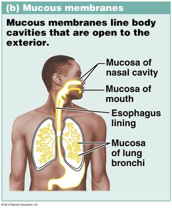
Mucous membranes (or mucosae) line body cavities and passages that open to the exterior environment, such as the respiratory, digestive, urinary, and reproductive tracts. They consist of an epithelial layer overlying a loose connective tissue layer called the lamina propria.
Epithelium: Varies by location (e.g., pseudostratified ciliated columnar in the respiratory tract, simple columnar in the digestive tract).
Lamina propria: Loose areolar connective tissue supporting the epithelium.
Mucus production: Goblet cells and glands secrete mucus, keeping the membrane moist and trapping pathogens and debris.
Examples of mucous membrane locations include the nasal cavity, mouth, esophagus, and bronchi.
Serous Membranes
Structure, Types, and Functions
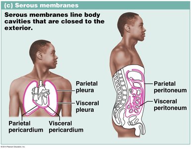
Serous membranes (serosae) line closed body cavities and cover the organs within them. They are composed of a simple squamous epithelium (mesothelium) overlying loose areolar connective tissue. The epithelium produces a slippery serous fluid that reduces friction between moving organs.
Visceral layer: Closest to the organ.
Parietal layer: Lines the cavity wall.
Serous fluid: Fills the space between the two layers, minimizing friction.
Major serous membranes include the pericardium (heart), pleura (lungs), and peritoneum (abdominopelvic organs).
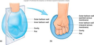
Balloon Analogy for Serous Membranes
The arrangement of serous membranes can be visualized using a balloon analogy: if a fist pushes into a balloon, the inner balloon wall represents the visceral layer, the outer wall the parietal layer, and the space between is the serous cavity filled with fluid.
Serous Membrane Types and Clinical Relevance
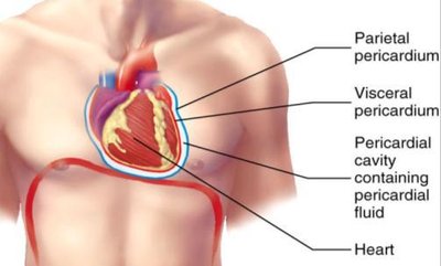
Pericardium (Heart)
The pericardium is the serous membrane surrounding the heart. It consists of the visceral pericardium (on the heart surface) and the parietal pericardium (lining the pericardial cavity). The pericardial cavity contains pericardial fluid, which reduces friction during heartbeats.
Pericarditis: Inflammation of the pericardium, often causing chest pain and impaired heart function.
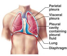
Pleura (Lungs)
The pleura is the serous membrane lining the thoracic cavity and covering the lungs. It consists of the visceral pleura (on the lung surface) and the parietal pleura (lining the thoracic wall). The pleural cavity contains pleural fluid, which reduces friction during breathing.
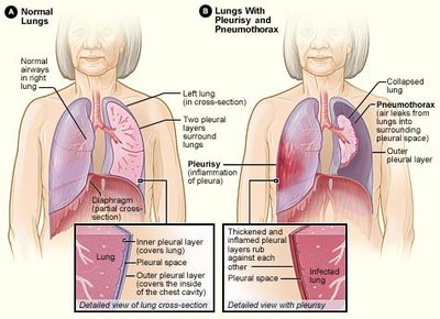
Pleurisy: Inflammation of the pleura, causing sharp chest pain with breathing.
Pneumothorax: Air or blood enters the pleural cavity, potentially causing lung collapse.
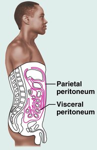
Peritoneum (Abdominopelvic Cavity)
The peritoneum lines the abdominopelvic cavity and covers the abdominal organs. The visceral peritoneum covers the organs, while the parietal peritoneum lines the cavity wall and diaphragm. The peritoneal cavity contains peritoneal fluid, which lubricates the organs.
Peritonitis: Inflammation of the peritoneum, often due to infection or injury.
Comparison: Mucous vs. Serous Membranes
Structural and Functional Differences
Feature | Mucous Membrane | Serous Membrane |
|---|---|---|
Location | Lines cavities open to exterior (e.g., respiratory, digestive tracts) | Lines closed cavities (e.g., thoracic, abdominal cavities) |
Epithelium | Varies (often columnar or pseudostratified) | Simple squamous (mesothelium) |
Connective Tissue | Lamina propria (areolar) | Areolar connective tissue |
Secretion | Mucus | Serous fluid |
Main Function | Protection, moistening, trapping debris | Lubrication, reducing friction |
Definition of a Membrane
A membrane is best described as a structure formed of an epithelial layer and a connective tissue layer that can be found both lining the lumen (inside) of an organ and covering the outside of an organ.
This definition encompasses both mucous and serous membranes, as well as the cutaneous membrane (skin).
Correct answer: C. A structure formed of an epithelial and connective tissue layer that could be found lining the lumen of an organ and covering the outside of an organ.
