 Back
Microscopic Anatomy and Organization of Skeletal Muscle
Study Guide - Smart Notes
 Tailored notes based on your materials, expanded with key definitions, examples, and context.
Microscopic Anatomy and Organization of Skeletal Muscle
Introduction to Skeletal Muscle Structure
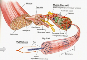
Skeletal muscle is a highly organized tissue responsible for voluntary movements in the human body. Its structure is hierarchical, with several levels of organization from the whole muscle down to the molecular components responsible for contraction.
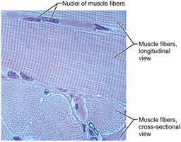
Skeletal muscle cells are also called muscle fibers.
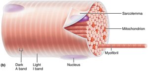
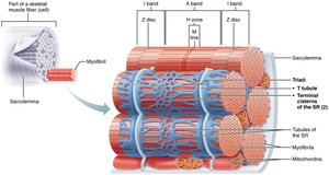
The plasma membrane of a muscle fiber is termed the sarcolemma.
Myofibrils are the contractile elements within muscle fibers, composed of actin and myosin filaments.
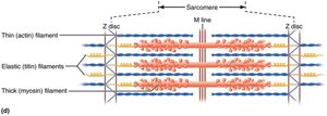
The sarcomere is the fundamental contractile unit, extending from one Z disc to the next.
Sarcoplasmic reticulum is a modified smooth endoplasmic reticulum that stores calcium ions necessary for muscle contraction.
Muscle Fiber and Sarcomere Structure
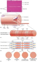
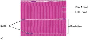
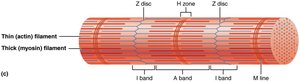
Each muscle fiber contains numerous myofibrils, which are further divided into repeating units called sarcomeres. The arrangement of actin (thin) and myosin (thick) filaments within the sarcomere gives skeletal muscle its striated appearance.
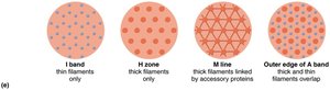
A bands (dark bands): Regions containing thick myosin filaments.
I bands (light bands): Regions containing thin actin filaments only.
Z discs: Define the boundaries of each sarcomere.
H zone: Central region of the A band where only myosin is present.
M line: Center of the H zone, where supporting proteins hold thick filaments together.
Connective Tissue Sheaths of Skeletal Muscle
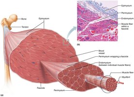
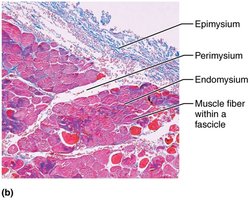
Skeletal muscle is supported and organized by three layers of connective tissue sheaths:
Epimysium: Surrounds the entire muscle.
Perimysium: Surrounds bundles of muscle fibers called fascicles.
Endomysium: Surrounds individual muscle fibers, lying just outside the sarcolemma.
Neuromuscular Junction
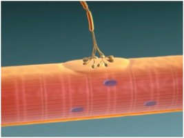
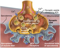
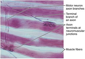
The neuromuscular junction (NMJ) is the specialized synapse where a motor neuron communicates with a skeletal muscle fiber to initiate contraction. This junction is essential for translating neural signals into muscle action.
Motor end plate: Specialized region of the sarcolemma with receptors for the neurotransmitter acetylcholine (ACh).
Synaptic cleft: The small gap between the axon terminal and the muscle fiber.
Acetylcholine (ACh): Neurotransmitter released from the motor neuron to stimulate muscle contraction.
Muscle Contraction: Overview
Muscle contraction is a complex process involving the interaction of actin and myosin filaments within the sarcomere, triggered by neural stimulation at the neuromuscular junction. The sliding filament theory explains how muscles generate force and shorten.
Isotonic contraction: Muscle changes length (shortens or lengthens) while the tension remains constant.
Isometric contraction: Muscle tension increases, but the muscle does not change length.
Steps in Muscle Contraction:
Action potential arrives at the neuromuscular junction.
Acetylcholine is released and binds to receptors on the sarcolemma.
Depolarization of the sarcolemma triggers calcium release from the sarcoplasmic reticulum.
Calcium binds to troponin, causing tropomyosin to move and expose binding sites on actin.
Myosin heads bind to actin, forming cross-bridges and pulling actin filaments toward the center of the sarcomere (power stroke).
ATP binds to myosin, causing it to detach from actin and reset for another cycle.
Key Equation:
The force generated by a muscle is proportional to the number of cross-bridges formed:
Practice and Learning Objectives
Identify the three types of muscle tissue (skeletal, cardiac, smooth).
Recognize the structures of a muscle fiber and the bands of a sarcomere.
Describe the structure and function of the neuromuscular junction.
List the steps in muscle contraction.
Distinguish between isotonic and isometric contractions.
Summary Table: Key Structures in Skeletal Muscle
Structure | Description | Function |
|---|---|---|
Epimysium | Outer connective tissue sheath | Surrounds entire muscle |
Perimysium | Connective tissue around fascicles | Bundles muscle fibers into fascicles |
Endomysium | Delicate connective tissue | Surrounds individual muscle fibers |
Sarcolemma | Plasma membrane of muscle fiber | Conducts action potentials |
Sarcoplasmic reticulum | Modified smooth ER | Stores and releases calcium ions |
Myofibril | Rod-like contractile element | Contains sarcomeres for contraction |
Sarcomere | Segment between two Z discs | Basic contractile unit |
Additional info: The notes above expand on the provided content with academic context, including definitions, explanations, and a summary table for clarity.
