 Back
BackMicroscopic Anatomy of the Stomach: Layers and Gastric Glands
Study Guide - Smart Notes
 Tailored notes based on your materials, expanded with key definitions, examples, and context.
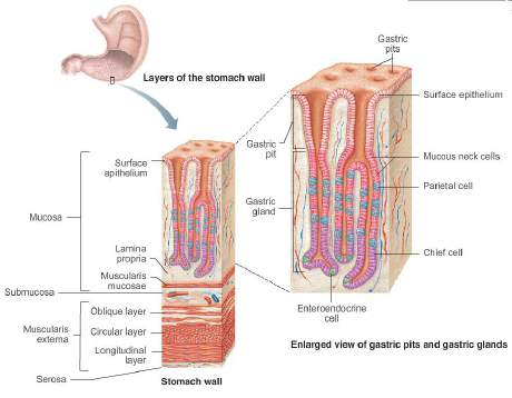
Q1. Label the terms on the figure of the stomach wall and gastric glands.
Background
Topic: Microscopic Anatomy of the Stomach
This question is testing your understanding of the structural organization of the stomach wall, including its layers and the specialized cells found in the gastric glands. This is a foundational concept in anatomy and physiology, especially for understanding digestive processes and the roles of different cell types in secretion and protection.
Key Terms and Concepts:
Mucosa: The innermost layer, containing the epithelium, lamina propria, and muscularis mucosae.
Submucosa: Connective tissue layer with blood vessels, nerves, and glands.
Muscularis externa: Muscle layer responsible for churning and mixing; includes oblique, circular, and longitudinal layers in the stomach.
Serosa: Outermost connective tissue covering.
Gastric pits and glands: Invaginations in the mucosa leading to glands that secrete gastric juice.
Cell types: Surface epithelium, mucous neck cells, parietal cells, chief cells, enteroendocrine cells.
Step-by-Step Guidance
Start by identifying the four main layers of the stomach wall from the innermost to the outermost: mucosa, submucosa, muscularis externa, and serosa. Note the unique presence of three muscle layers in the muscularis externa of the stomach (oblique, circular, longitudinal).
Within the mucosa, locate the surface epithelium, lamina propria, and muscularis mucosae. The surface epithelium is made up of simple columnar cells that secrete mucus for protection.
Observe the gastric pits, which are invaginations of the mucosa leading to the deeper gastric glands. These glands contain several specialized cell types.
Identify the main cell types in the gastric glands: mucous neck cells (secrete mucus), parietal cells (secrete HCl and intrinsic factor), chief cells (secrete pepsinogen), and enteroendocrine cells (release hormones).
Match each label on the diagram to the correct structure or cell type based on their location and function. For example, parietal cells are typically found in the middle region of the gland, while chief cells are deeper.
Try solving on your own before revealing the answer!
Final Answer:
The correct labels for the figure are as follows:
Surface epithelium: Simple columnar cells lining the lumen.
Gastric pit: Invagination leading to the gland.
Mucous neck cells: Secrete acidic mucus, found in the neck region of the gland.
Parietal cell: Secretes HCl and intrinsic factor, located in the middle region.
Chief cell: Secretes pepsinogen, found deeper in the gland.
Enteroendocrine cell: Secretes hormones into the lamina propria.
Lamina propria: Connective tissue supporting the epithelium.
Muscularis mucosae: Thin muscle layer beneath the mucosa.
Oblique, circular, longitudinal layers: Three muscle layers of the muscularis externa.
Serosa: Outermost connective tissue covering.
This organization allows the stomach to perform its digestive and protective functions efficiently.
