 Back
Midterm Lab Study Guide: Human Body Orientation, Tissues, and Cells
Study Guide - Smart Notes
 Tailored notes based on your materials, expanded with key definitions, examples, and context.
Body Organization and Anatomical Terminology
Body Planes
Understanding anatomical planes is essential for describing locations and movements of body parts. The three primary planes are:
Frontal (Coronal) Plane: Divides the body into anterior (front) and posterior (back) sections.
Transverse (Horizontal) Plane: Divides the body into superior (upper) and inferior (lower) parts.
Median (Midsagittal) Plane: Divides the body into equal right and left halves.
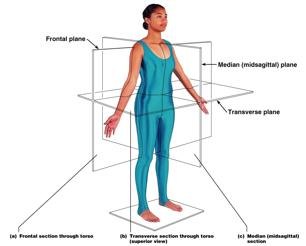
Example: A CT scan often uses the transverse plane to create cross-sectional images of the body.
Anatomical Terms: Anterior and Posterior Views
Precise anatomical terminology is used to describe locations and regions of the human body. The anterior (front) and posterior (back) views label key surface landmarks.
Anterior (Ventral): Refers to the front of the body (e.g., chest, abdomen, face).
Posterior (Dorsal): Refers to the back of the body (e.g., back, buttocks).
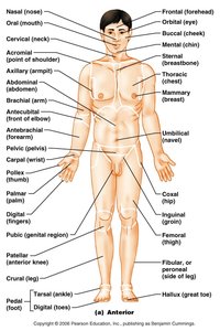
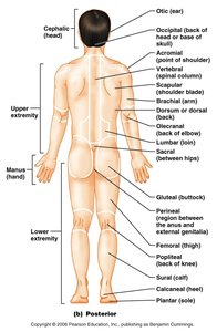
Example: The sternum is anterior to the heart, while the scapula is posterior to the rib cage.
Abdominopelvic Regions and Quadrants
The abdomen is divided into regions and quadrants for clinical and anatomical reference. The nine-region method is commonly used to localize organs and pain.
Regions: Right/Left hypochondriac, epigastric, right/left lumbar, umbilical, right/left iliac (inguinal), hypogastric (pubic).
Quadrants: Right upper, left upper, right lower, left lower (not shown in image).
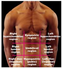
Example: The appendix is typically located in the right iliac (inguinal) region.
Tissues: The Living Fabric
Classification of Tissues
All tissues are classified into four basic types:
Epithelial Tissue: Covers surfaces, lines cavities, forms glands.
Connective Tissue: Supports, binds, and protects organs.
Muscle Tissue: Responsible for movement.
Nervous Tissue: Initiates and transmits electrical impulses.
Example: The skin contains all four tissue types in its structure.
Epithelial Tissue Types
Epithelial tissues are classified by cell shape and number of layers:
Simple: One cell layer thick.
Stratified: Multiple cell layers.
Squamous: Flat cells.
Cuboidal: Cube-shaped cells.
Columnar: Tall, column-like cells.
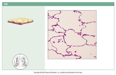
Simple Squamous Epithelium
Single layer of flat cells; allows for rapid diffusion and filtration. Found in air sacs of lungs and lining of blood vessels.
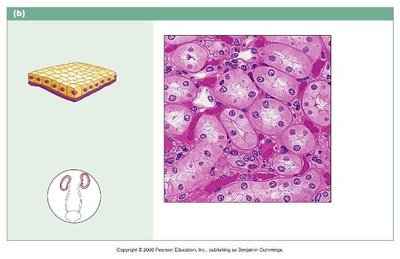
Simple Cuboidal Epithelium
Single layer of cube-shaped cells; functions in secretion and absorption. Found in kidney tubules and glandular ducts.
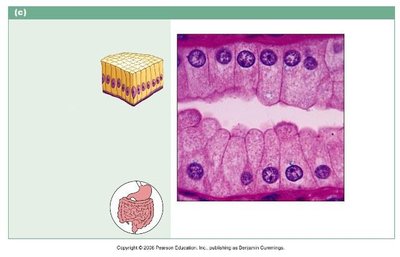
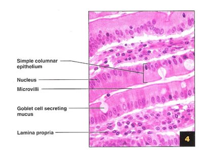
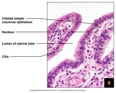
Simple Columnar Epithelium (Non-ciliated and Ciliated)
Single layer of tall, column-like cells; may have microvilli or cilia. Non-ciliated type lines most of the digestive tract; ciliated type lines small bronchi and uterine tubes.
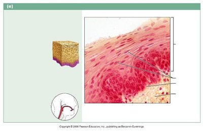
Stratified Squamous Epithelium
Multiple layers of flat cells; protects underlying tissues in areas subject to abrasion. Found in the skin, mouth, and esophagus.
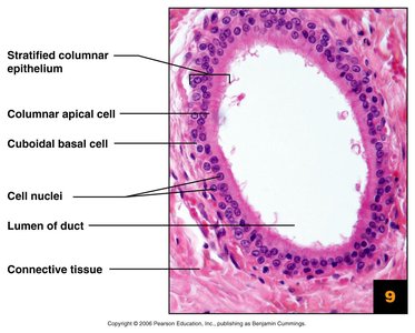
Stratified Columnar Epithelium
Rare; consists of several layers with columnar cells on the surface. Found in some ducts and glands.
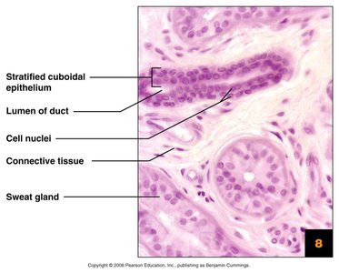
Stratified Cuboidal Epithelium
Typically two layers of cube-shaped cells; found in sweat gland ducts and mammary glands.
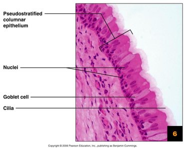
Pseudostratified Columnar Epithelium (Ciliated)
Appears stratified but is a single layer with nuclei at different levels; often ciliated. Lines the respiratory tract.
Connective Tissue Types
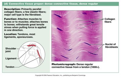
Dense Regular Connective Tissue
Composed of parallel collagen fibers; provides strong attachment between structures. Found in tendons and ligaments.
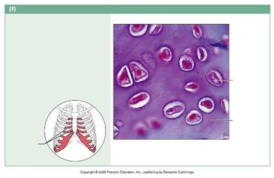
Hyaline Cartilage
Most common cartilage; provides support with some flexibility. Found in the nose, trachea, and at the ends of long bones.
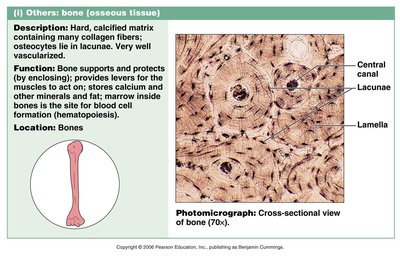
Bone Tissue (Compact and Spongy)
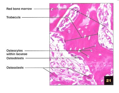
Bones are composed of compact and spongy tissue. Compact bone is dense and forms the outer layer; spongy bone is lighter and found inside bones.
Blood (A Connective Tissue)
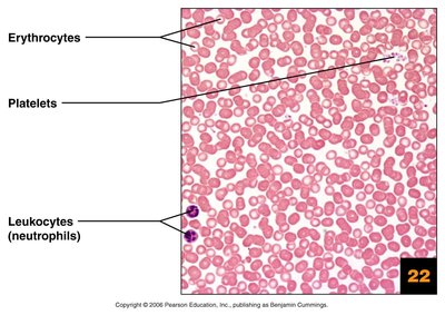
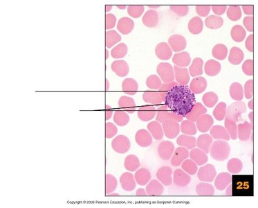
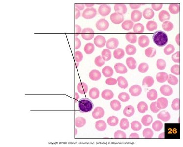
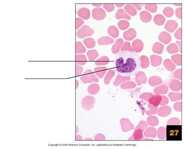
Blood is a fluid connective tissue composed of erythrocytes (red blood cells), leukocytes (white blood cells), and platelets suspended in plasma.
White Blood Cells (Leukocytes)
There are several types of leukocytes, each with distinct functions in immunity:
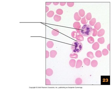
Neutrophils: Phagocytize bacteria.
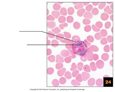
Eosinophils: Combat parasites and allergens.
Basophils: Release histamine in allergic reactions.
Lymphocytes: Produce antibodies and regulate immune response.
Monocytes: Differentiate into macrophages for phagocytosis.
Adipose Tissue
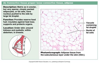
Adipose tissue is a loose connective tissue that stores fat, insulates the body, and protects organs.
Muscle and Nervous Tissue
Muscle tissue is specialized for contraction and movement, while nervous tissue is specialized for communication via electrical impulses.
Muscle Tissue Types: Skeletal (voluntary movement), cardiac (heart), and smooth (walls of hollow organs).
Nervous Tissue: Composed of neurons (transmit signals) and neuroglial cells (support neurons).
Example: Skeletal muscle enables limb movement, while neurons transmit sensory information to the brain.
Additional info: For lab exams, focus on identifying and labeling all structures indicated by leader lines in diagrams and slides. Use available resources for practice and clarification.
